Dental
Showing 25–36 of 37 results
-
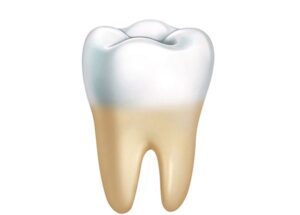
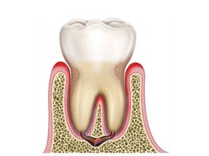
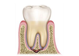
Molar tooth anatomy
24,50 € Add to basketMolar tooth anatomy is a posterior tooth specialized in
grinding food. Its structure includes enamel, dentin,
and pulp, as well as roots that attach it to the bone
via the periodontal ligament.


-
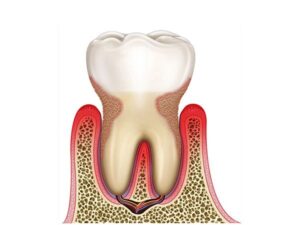
Periodontal disease
24,50 € Add to basketPeriodontal disease is a progressive condition affecting the gums and
supporting tissues, starting as gingivitis and potentially leading to
bone loss and tooth mobility if untreated. -
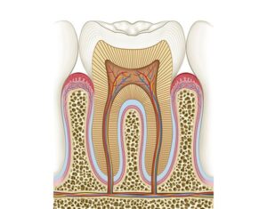
Physiology of the molar
24,50 € Add to basketMolars have a multilayer structure including enamel,
dentin and pulp. Their crowns grind food while roots
anchor the tooth to the jaw through the periodontal
ligament. -
Stage II periodontitis
24,50 € Add to basketThe stage II periodontitis has an altered function in dental
support. Moreover, under normal conditions, the gums and
bone keep the tooth stable. Additionally, the periodontal
ligament distributes chewing forces. -
Stages of periodontitis
24,50 € Add to basketThe stages of periodontitis have a general function in disease
classification. Moreover, under normal conditions, gums and
bone maintain tooth stability. Additionally, the periodontal
ligament distributes forces. Furthermore, they ensure oral health. -
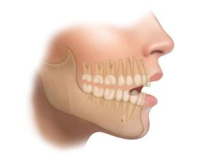
Teeth and jaw
24,50 € Add to basketDentition and the mandible work together in the
stomatognathic system to support teeth and allow
proper chewing and oral function. -
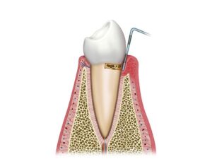
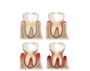
The periodontal initial phase
24,50 € Add to basketPeriodontal initial phase. The illustration regarding this phase
describes the conditions that impact the gums and supporting
structures. The onset manifests as gingivitis with inflammation
and bleeding, potentially evolving into periodontitis with bone
loss due to lack of oral hygiene. -
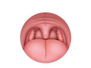
The tonsils
24,50 € Add to basketThe tonsils are lymphoid organs located in the throat
that help protect the body against bacteria and viruses.
They are part of the immune system and participate in
the defense against infections. -
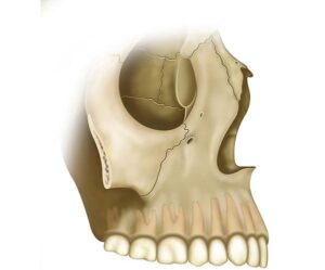
The upper jaw
24,50 € Add to basketThe maxilla is a craniofacial bone that supports the
upper teeth and contributes to the structure of the
palate, nasal cavity, and eye orbit. -
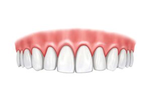
The upper teeth
24,50 € Add to basketUpper dentition includes incisors, canines, premolars,
and molars located in the maxilla. These teeth are essential
for chewing, speech, and facial appearance. -
Tooth attachment
24,50 € Add to basketTooth attachment depends on the periodontium, a set of
tissues that connect the tooth root to the alveolar bone.
It includes the periodontal ligament, root cementum,
alveolar bone, and gums. -
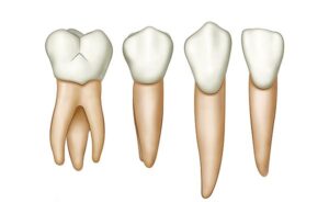
Types of teeth
24,50 € Add to basketTeeth are classified into incisors, canines, premolars,
and molars. Each type performs specific functions in
cutting, tearing, and grinding food during chewing.