Melanoma
Showing 13–24 of 53 results
-
Facial dermatomyositis
24,50 € Add to basketFacial dermatomyositis. This graphic representation details the
skin alterations and inflammatory symptoms characteristic of this
autoimmune condition. The content describes the erythema and
muscular involvement in the facial region to facilitate precise clinical
identification.


-
Fingers with dermatomyositis
24,50 € Add to basketFingers with dermatomyositis: this anatomical illustration
highlights the impacts of this autoimmune disease on the limbs.
The condition causes muscle inflammation and skin rashes such
as Gottron’s papules. Early diagnosis is essential to treat weakness
and characteristic visual changes. -
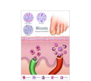
Fungal skin infection
24,50 € Add to basketFungal skin infection. This schematic illustrates how fungi affect
the skin and keratinized tissues in various body areas. The content
details symptoms of itching and scaling, emphasizing the importance
of hygiene and the use of antifungals for effective management. -
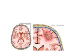
Glioblastoma brain tumor
24,50 € Add to basketGlioblastoma brain tumor. This realistic illustration shows a coronal
section of the brain affected by a highly aggressive glioblastoma. The
content describes its invasive nature and highlights the importance of
this visual resource for diagnosis and education about the most lethal
brain tumor. -
Glioblastoma multiforme tumor
24,50 € Add to basketGlioblastoma multiforme tumor. This illustration details the
cross-section of the brain affected by a highly aggressive glioblastoma
multiforme. The content describes its infiltrative growth in the cerebral
hemispheres and highlights the importance of this visual resource for
clinical diagnosis, medical education. -
Hand with dermatomyositis
24,50 € Add to basketThis representation of a hand with dermatomyositis illustrates
Gottron’s papules and skin inflammation. Learn about key symptoms,
from muscle weakness to rashes, and the importance of early diagnosis
to improve the quality of life through specialized medical treatments
and therapies. -
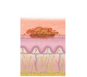
Impetigo crust
24,50 € Add to basketImpetigo crust. This visual representation details the honey-colored
and yellowish lesions caused by bacterial infections of staphylococci or
streptococci. The content explains the distinctive appearance of the
infection and highlights the importance of hygiene and antibiotics
treatment for rapid healing. -
Impetigo skin infection
24,50 € Add to basketImpetigo skin infection. This diagram details the formation of
vesicles and yellowish crusts caused by streptococci or staphylococci.
The content technically describes the evolution of the lesions and
highlights the importance of antibiotic treatment and hygiene for their
resolution. -
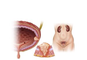
Malignant bladder tumor
24,50 € Add to basketMalignant bladder tumor. This anatomical illustration shows
the human urinary system and a bladder with tumor growth. The
content highlights the importance of this visual resource for clinical
diagnosis of neoplasia on urinary function. -
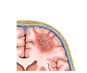
Malignant brain tumor
24,50 € Add to basketMalignant brain tumor. This coronal section illustration details
the invasion of a malignant neoplasm in the brain parenchyma and
its effect on anatomy. The content highlights the importance of
technical visualization for accurate diagnosis. -
Malignant glioblastoma tumor
24,50 € Add to basketMalignant glioblastoma tumor. This realistic illustration of a
cerebral cross-section details the invasive growth of glioblastoma
multiforme (GBM). The content describes the aggressiveness of this
primary brain tumor and highlights the importance of visualization. -
Malignant thyroid tumor
24,50 € Add to basketMalignant thyroid tumor. This anatomical illustration details the
location of a malignant neoplasm in the thyroid gland and its relationship
to the trachea. The content highlights the utility of this visual resource for
clinical diagnosis