Physiology
Showing 13–16 of 16 results
-
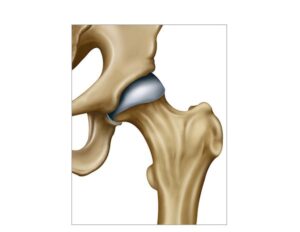
The coxofemoral joint
24,50 € Add to basketThe hip joint. This anatomical union between the acetabulum
and the femur facilitates stability and body weight support. Its
function allows for flexion and rotation movements indispensable
for walking, running, and maintaining proper posture during
daily activities.


-
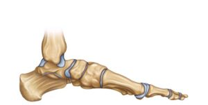
The foot lateral view
24,50 € Add to basketThe foot lateral view. An anatomical representation of the lower
limb detailing the connection between the tibia, fibula, and tarsal
bones. The image shows the talus, calcaneus, navicular, and cuboid.
elements that work together to provide structure, balance, and
mobility during human gait. -
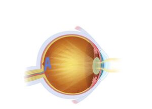
The vision with cataracts
24,50 € Add to basketVision with cataracts. The scheme reflects the opacity of the lens, as
the natural lens of the eye, which blurs the sight and affects correct
focusing. The surgical treatment consists of the extraction of the
opaque tissue and its replacement with an artificial lens to restore
visual clarity. -
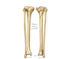
Tibia and fibula anatomy
24,50 € Add to basketTibia and fibula anatomy. These two long bones form the skeletal
structure of the lower leg. The tibia stands out for its ability to support
body weight and transmit loads, while the fibula provides lateral stability
and serves as an anchor for muscles essential for human locomotion.