Joint
Showing 1–12 of 13 results
-
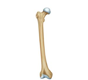
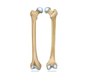
Anatomy of the femur bone
24,50 € Add to basketAnatomy of the femur bone. The longest and strongest bone in
the body is located between the hip and the knee. Its structure,
composed of head, neck, and shaft, facilitates body weight support
and musculoskeletal mobility through its connection with the pelvis
and the tibia.


-
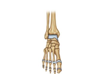
Frontal view of the foot
24,50 € Add to basketFrontal view of the foot. Technical representation of the bony
structures, including phalanges, metatarsals, and tarsal bones.
The image details the organization of the talus, calcaneus, and
cuneiforms, elements that provide the necessary anatomical base
for balance and locomotion. -
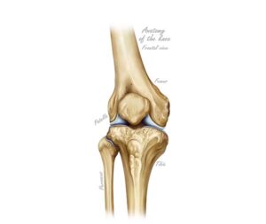
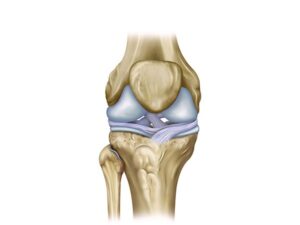
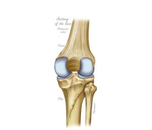
Frontal view of the knee
24,50 € Add to basketFrontal view of the knee. Technical illustration of the joint showing
the arrangement of the femur, tibia, fibula, and patella. The image
displays the articular surfaces and cartilage, key elements for flexion
and human locomotion in the activities of daily living. -
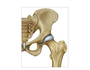
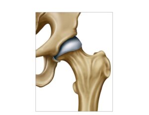
Hip and femur joint
24,50 € Add to basketHip and femur joint. The anatomical union between the acetabulum
and the femoral head allows the execution of flexion, extension, and
rotation movements. This bony structure supports the weight of the
torso and facilitates human locomotion, being vital for stability and
physical balance. -
Knee cruciate ligament
24,50 € Add to basketKnee cruciate ligament. This anatomical structure joins the femur
with the tibia to provide internal stability. The anterior and posterior
cruciate ligaments are fundamental to preventing excessive bone
displacement, facilitating the diagnosis and treatment of common
joint injuries. -
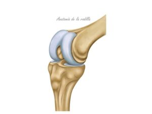
Knee function
24,50 € Add to basketKnee function. This joint acts as a complex hinge that facilitates
the flexion and extension of the leg. Its structure, composed of the
femur and tibia, supports body weight and guarantees the stability
necessary to walk, run, and perform daily activities safely. -
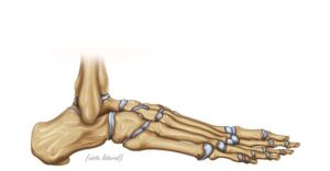
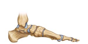
Lateral view of the foot
24,50 € Add to basketLateral view of the foot. Illustration of the bony anatomy showing
the connection of the tibia and fibula with the talus and calcaneus.
The image details the tarsal bones, such as the navicular and cuboid,
essential for maintaining balance, arch structure, and human mobility. -
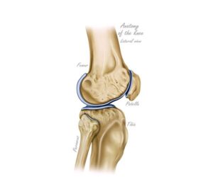
Lateral view of the knee
24,50 € Add to basketLateral view of the knee. This anatomical perspective details the
relationship between the femur, tibia, and patella. The illustration
highlights the articular surfaces and cartilages that allow for flexion
and extension of the leg, being fundamental for understanding
locomotion and skeletal stability. -
Morphology of the femur bone
24,50 € Add to basketMorphology of the femur bone. The longest bone in the thigh
presents a robust structure with a well-defined head, neck, and body.
Its frontal and posterior views reveal details such as the condyles and
the intertrochanteric crest, crucial elements for body support and
joint connection. -
Posterior view of the knee
24,50 € Add to basketPosterior view of the knee. Technical illustration detailing the
back of the joint, including the femur, tibia, and fibula. The image
shows the structures necessary for flexion and leg biomechanics,
serving as an essential tool for evaluating functions and injuries. -
The coxofemoral joint
24,50 € Add to basketThe hip joint. This anatomical union between the acetabulum
and the femur facilitates stability and body weight support. Its
function allows for flexion and rotation movements indispensable
for walking, running, and maintaining proper posture during
daily activities. -
The foot lateral view
24,50 € Add to basketThe foot lateral view. An anatomical representation of the lower
limb detailing the connection between the tibia, fibula, and tarsal
bones. The image shows the talus, calcaneus, navicular, and cuboid.
elements that work together to provide structure, balance, and
mobility during human gait.