Voice
Showing 1–12 of 18 results
-
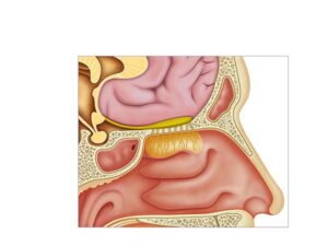
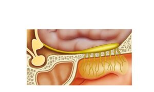
Anatomy and physiology of smell
24,50 € Add to basketAnatomy and physiology of smell: a system that detects odors
in the nasal mucosa, it transmits signals through the olfactory
nerve and processes them in the olfactory bulb in the brain.


-
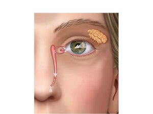
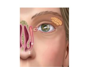
Anatomy of the lacrimal glands
24,50 € Add to basketAnatomy of the lacrimal glands: eye structures that produce
tears, lubricate the surface, remove particles, and protect
against infections, maintaining a balance between production
and drainage. -
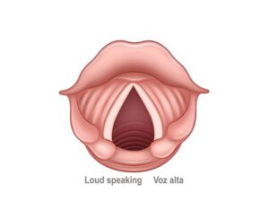
Forced phonation
24,50 € Add to basketForced phonation: excessive voice use increases pressure
on the vocal cords, leading to fatigue, irritation, or injury,
affecting voice quality without proper vocal techniques. -
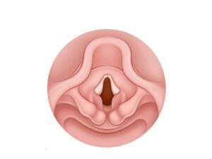
Infant vocal cords
24,50 € Add to basketInfant vocal cords: small, delicate structures in the larynx
that allow sound production, crying, and early language
development in babies. -
Lacrimal glands
24,50 € Add to basketLacrimal glands: structures in the upper outer eye that produce
tears, lubricate the ocular surface, remove particles, and protect
against infections with substances like antibodies. -
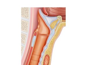
Larynx anatomy
24,50 € Add to basketLarynx anatomy: an organ of the respiratory system with vocal
cords, cartilages, and epiglottis that regulate airflow, produce voice,
and protect the airways. -
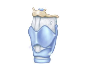
Larynx anatomy
24,50 € Add to basketLarynx anatomy: a cartilaginous organ in the neck that
regulates airflow, houses the vocal cords for phonation,
and protects airways through the epiglottis. -
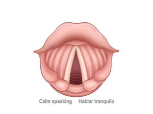
Normal phonation
24,50 € Add to basketNormal phonation: a process where the vocal cords vibrate as air
passes from the lungs, producing sound and allowing modulation
of pitch, volume, and intonation in speech. -
Olfactory bulb anatomy
24,50 € Add to basketOlfactory bulb anatomy: a structure at the base of the brain
that receives signals from the olfactory mucosa via the olfactory
nerve and processes odor information. -
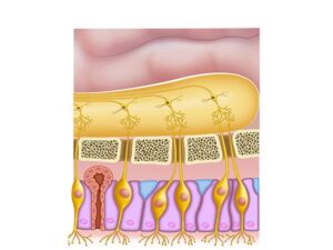
Olfactory nerve diagram
24,50 € Add to basketOlfactory nerve diagram: the first cranial nerve that carries
signals from the olfactory mucosa to the olfactory bulb, enabling
odor perception and identification. -
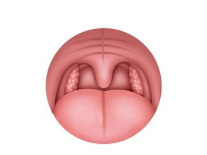
Palatine tonsils
24,50 € Add to basketPalatine tonsils: lymphoid organs in the oropharynx that
are part of the immune system, filtering bacteria and viruses
and helping protect the body from infections. -
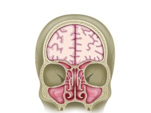
Paranasal sinuses coronal plane
24,50 € Add to basketParanasal sinuses in the coronal plane: shows the frontal sinus
in the forehead, its connection to the nasal cavity, and its roles
in voice resonance, skull protection, and air conditioning.