Hearing
Showing 1–12 of 14 results
-
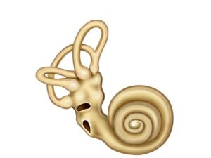
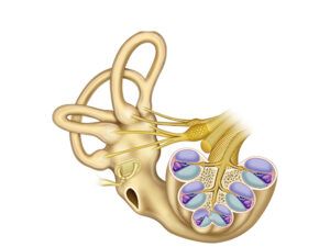
Anatomy of the cochlea
24,50 € Add to basketThe anatomy of the cochlea shows a spiral structure
in the inner ear that contains the organ of Corti. Hair
cells convert sound vibrations into electrical signals,
while fluid-filled chambers respond to different sound
frequencies.


-
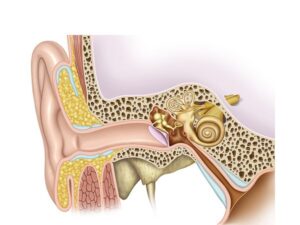
Anatomy of the ear
24,50 € Add to basketThe anatomy of the ear includes the outer, middle, and
inner ear. These structures work together to capture sound,
transmit vibrations, and maintain balance. -
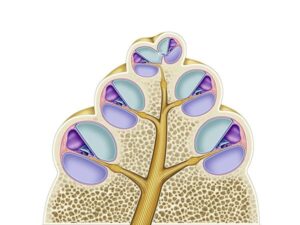
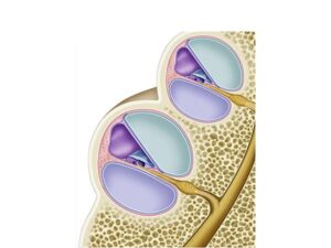
Cochlea cross section
24,50 € Add to basketThe cross-section of the cochlea shows the spiral structure
of the inner ear. It contains the organ of Corti, where hair
cells transform sound vibrations into electrical signals that
allow us to perceive different frequencies and tones. -
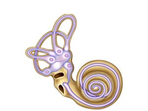
Cochlear ducts
24,50 € Add to basketCochlear ducts are spiral structures inside the cochlea of
the inner ear. They contain fluid and the organ of Corti,
where hair cells convert sound vibrations into electrical
signals that allow the brain to detect different sound
frequencies. -
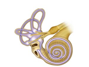
Cochlear vestibular system
24,50 € Add to basketThe vestibulocochlear system connects the cochlea and
the vestibular system with the brain. The cochlea processes
sound, while the vestibular system controls balance and
spatial orientation. -
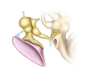
Eardrum and Cochlea
24,50 € Add to basketThe eardrum and cochlea work together in the hearing
process. The eardrum captures sound waves and sends
vibrations to the inner ear, where the cochlea converts
them into electrical signals interpreted by the brain. -
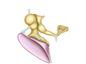
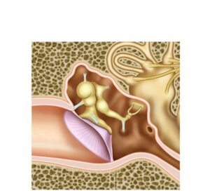
Eardrum and ossicles
24,50 € Add to basketThe tympanic membrane and auditory ossicles work
together to transmit and amplify sound waves. The
malleus, incus, and stapes carry vibrations to the inner
ear, where they become electrical signals interpreted
by the brain. -
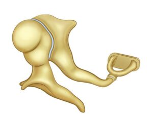
Middle ear bones
24,50 € Add to basketMiddle ear ossicles, the malleus, incus, and stapes,
transmit and amplify sound vibrations from the eardrum
to the inner ear. This process allows sound waves to be
converted into electrical signals that the brain interprets
as sound. -
Middle ear structure
24,50 € Add to basketThe middle ear is located in the tympanic cavity and
contains the eardrum and auditory ossicles. These
structures transmit and amplify sound vibrations toward
the inner ear, allowing efficient hearing and proper
sound processing. -
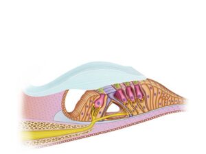
Organ of Corti
24,50 € Add to basketThe organ of Corti is located in the cochlea of the
inner ear. It contains hair cells that convert sound
vibrations into electrical signals transmitted to the
brain through the auditory nerve, allowing the
perception of different frequencies and tones. -
Physiology of the cochlea
24,50 € Add to basketThe physiology of the cochlea explains how sound
vibrations become electrical signals. In the organ of
Corti, hair cells detect vibrations and send impulses
through the auditory nerve to the brain for sound
interpretation. -
The Cochlear Duct
24,50 € Add to basketThe cochlear duct is located inside the cochlea in the
inner ear. It contains the organ of Corti, where hair cells
convert sound vibrations into electrical signals that allow
the brain to detect different frequencies and sounds.