Ocular
Showing 13–24 of 68 results
-
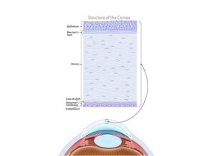
Diagram of the cornea
24,50 € Add to basketThe corneal layer is the transparent front structure of
the eye. It allows light to enter the eye, contributes to
light refraction, and protects internal eye structures,
helping form clear images on the retina.


-
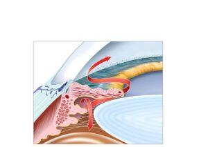
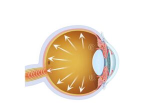
Drainage blockage in glaucoma
24,50 € Add to basketDrainage blockage in glaucoma shows how obstruction of the
aqueous humor flow increases intraocular pressure. This fluid
drains through the trabecular meshwork and Schlemm’s canal,
but when drainage is blocked, eye pressure rises progressively
damaging the optic nerve. -
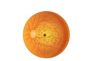
Dry Macular Degeneration
24,50 € Add to basketDry Macular Degeneration (AMD), dry form, illustrates how, in
this disease, the macula—the central part of the retina—gradually
deteriorates. As the macula loses cells, an accumulation of cellular
debris known as drusen forms. -
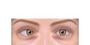
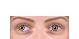
Esotropia strabismus
24,50 € Add to basketEsotropic strabismus is a form of strabismus where one eye
deviates inward compared to the other. This condition can result
in double vision and visual coordination problems. -
Exotropia strabismus
24,50 € Add to basketThe eye in strabismus hypertropia is a type of strabismus in which
one eye is deflected upwards compared to the other. This can cause
double vision and visual coordination problems. -
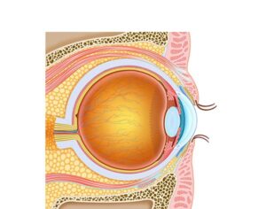
Eye and eyelids anatomy
24,50 € Add to basketEye and eyelid anatomy includes structures responsible
for vision and protection. The eye focuses light onto the
retina, while the eyelids protect the eyeball and distribute
tears to keep the ocular surface hydrated. -
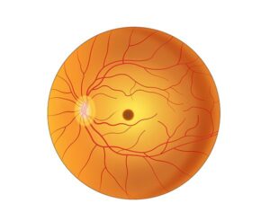
Eye with healthy macula
24,50 € Add to basketThe healthy macula is responsible for central and detailed
vision. Its photoreceptor cells allow clear perception of colors,
contrasts, and fine visual details, making it essential for tasks
such as reading and recognizing faces. -
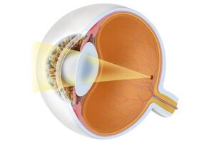
Eye with normal vision.
24,50 € Add to basketNormal vision occurs when light enters the eye through
the cornea, passes through the pupil, and is focused by the
lens onto the retina. Through accommodation, the eye can
adjust focus for near and distant objects, allowing clear
and sharp visual perception. -
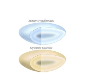
Healthy and cataract lens
24,50 € Add to basketA healthy eye lens and another affected by cataracts. In a healthy
eye, the lens is clear, allowing light to pass to the retina without
obstruction. In contrast, a lens with cataracts looks cloudy, making
vision difficult and causing blur. -
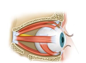
Human extraocular muscles
24,50 € Add to basketHuman extraocular muscles control the movement of the eyeball.
These six muscles work together to produce vertical, horizontal,
and rotational eye movements, allowing both eyes to coordinate
their position and maintain accurate visual tracking. -
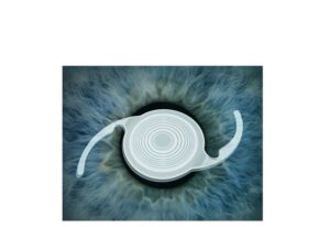
Intraocular lens
24,50 € Add to basketIntraocular lens. This artificial lens is implanted in the eye after the
removal of the natural lens during cataract surgery. The intraocular
lens replaces the cloudy lens, allowing light to focus correctly on the
retina and restoring vision. height=”228″ />=”https://ilusmedical.com/head-and-neck-disorders/?lang=en” target=”_blank” rel=”noopener”>