Bone
Showing 37–48 of 108 results
-
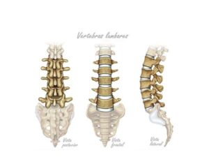
Lumbar vertebrae
24,50 € Add to basketLumbar vertebrae. The bony structures in the lower region of
the spine are shown here. These vertebrae, numbered from L1
to L5, support the weight of the torso and allow for movements
such as flexion and extension.

-
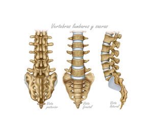
Lumbar vertebrae and sacrum
24,50 € Add to basketLumbar and sacral vertebrae. These show the bony structures
in the lower part of the spine. Numbered L1 through L5, the
lumbar vertebrae support the weight of the torso and allow for movements
such as flexion and extension. The sacrum, formed by fused vertebrae,
connects the lumbar spine to the pelvis.</p>
-
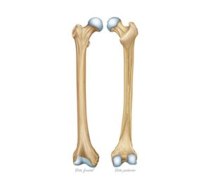
Morphology of the femur bone
24,50 € Add to basketMorphology of the femur bone. The longest bone in the thigh
presents a robust structure with a well-defined head, neck, and body.
Its frontal and posterior views reveal details such as the condyles and
the intertrochanteric crest, crucial elements for body support and
joint connection. -
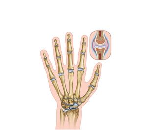
Morphology of the hand
24,50 € Add to basketAnatomy of the hand. This is a complex bony structure
that includes metacarpals, phalanges, and carpal bones.
The metacarpals form the base of the fingers, while the
phalanges make up the fingers themselves. -
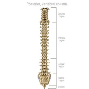
Posterior vertebral organization
24,50 € Add to basketPosterior view of the vertebral structure, highlighting its essential
function for mobility. Composed of cervical (C1-C7), thoracic (T1-T12),
lumbar (L1-L5), and sacral (S1-S5) vertebrae, and the coccyx, this
structure extends from the nape of the neck to the base of the back. -
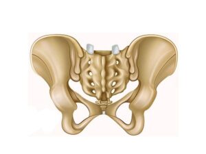
Posterior view of the hip
24,50 € Add to basketPosterior view of the hip. This vital joint connects the femur
to the sacrum, a triangular bone at the base of the spine. This
complex connection allows essential movements such as flexion
and rotation. -
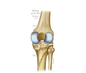
Posterior view of the knee
24,50 € Add to basketPosterior view of the knee. Technical illustration detailing the
back of the joint, including the femur, tibia, and fibula. The image
shows the structures necessary for flexion and leg biomechanics,
serving as an essential tool for evaluating functions and injuries. -
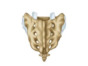
Posterior view of the sacrum
24,50 € Add to basketPosterior view of the sacrum: the triangular region at the bottom
of the spine. Numbered S1-S5, it features the median sacral crest,
intermediate and lateral crests, and four pairs of posterior sacral
foramina. -
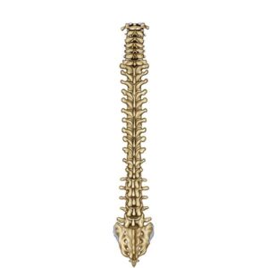
Posterior view of the spine
24,50 € Add to basketPosterior view of the spine. This structure is essential for mobility
and comprises the cervical vertebrae (C1–C7), thoracic (T1–T12),
lumbar (L1–L5), sacral (S1–S5), and coccyx. From the nape of the
neck to the lower back, it provides support and houses the spinal cord. -
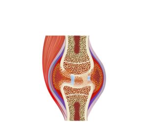
Rheumatoid arthritis
24,50 € Add to basketRheumatoid arthritis is an autoimmune disease causing chronic
inflammation and cartilage destruction. Moreover, it leads to pain,
stiffness, and joint deformity. Treatment includes disease-modifying
drugs, anti-inflammatory therapy, and physical rehabilitation. -
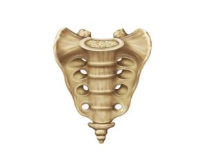
Sacrum frontal view
24,50 € Add to basketThe sacrum, viewed from the front, presents the fused triangular
structure at the base of the spine. Numbered S1-S5, the sacrum
forms the sacroiliac joint with the pelvis; the sacral crests and sacral
foramina are visible. -
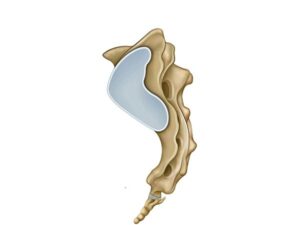
Sacrum lateral view
24,50 € Add to basketLateral view of the sacrum, with a triangular and fused structure
located at the base of the spine. Numbered S1-S5, the sacrum connects
to the pelvis and forms the sacroiliac joint. Lateral view of the sacrum.
The sacral crests and sacral foramina are visible.