Heart
Showing 85–96 of 108 results
-
The heart appendage
24,50 € Add to basketThe heart appendage has a general function related to atrial
dynamics. Moreover, under normal conditions, it contributes to
left atrial contraction. Additionally, it may serve as a temporary
blood reservoir.


-
The heart attack
24,50 € Add to basketThe heart attack has an altered function in myocardial perfusion.
Moreover, under normal conditions, the coronary arteries supply
oxygen-rich blood. Additionally, the heart maintains systemic
circulation. -
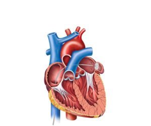
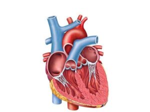
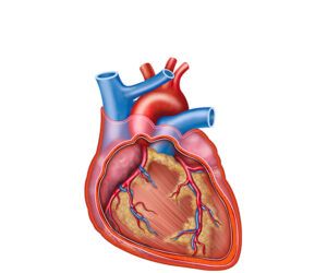
The heart in the sagittal plane
24,50 € Add to basketThe heart in the sagittal plane shows the heart divided
into right and left halves. This view highlights the atria,
ventricles, and heart valves, helping explain cardiac
anatomy and blood flow through the chambers. -
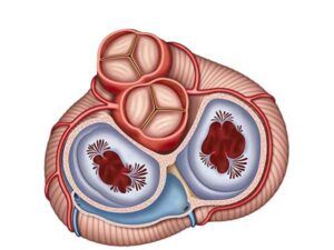
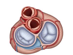
The heart valves
24,50 € Add to basketHeart valves regulate blood flow within the heart. The
mitral, tricuspid, aortic, and pulmonary valves open and
close during each heartbeat to ensure one-way blood flow
circulation through the heart and major arteries. -
The heart valves
24,50 € Add to basketHeart valves regulate blood flow inside the heart.
The mitral, tricuspid, aortic, and pulmonary valves
open and close during each heartbeat to maintain
one-way circulation. -
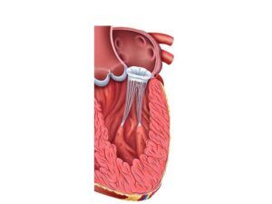
The mitral valve
24,50 € Add to basketThe mitral valve regulates blood flow between the left
atrium and the left ventricle. Its opening and closing during
the cardiac cycle ensure one-way blood circulation through
the heart. -
The pericarditis
24,50 € Add to basketThe pericarditis has an altered function in heart protection.
Moreover, under normal conditions, the pericardium reduces
friction during heartbeats. Additionally, it allows proper
cardiac movement. -
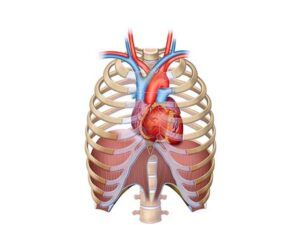
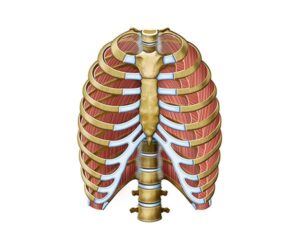
The rib cage
24,50 € Add to basketThe rib cage is a skeletal structure made of ribs,
sternum, and thoracic vertebrae. It protects the
heart and lungs while supporting breathing
movements. -
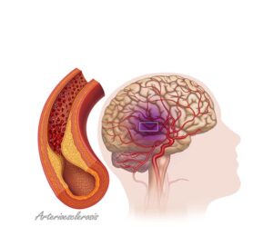
The stroke
24,50 € Add to basketThe stroke. A serious event caused by an obstruction or rupture
of brain vessels that disrupts blood flow, causing neurological
damage. Your immediate attention is key to reducing sequelae
such as loss of mobility, language, or functions. cognitive. -
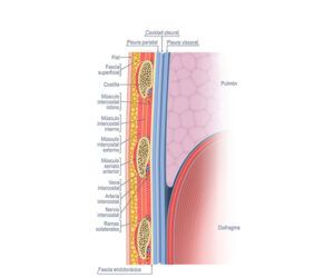
The thoracic cavity layers
24,50 € Add to basketThoracic cavity layers include skin, subcutaneous tissue,
intercostal muscles, ribs, and costal cartilage. Together, they
protect the heart and lungs while supporting breathing
movements. -
The thoracic cavity muscles
24,50 € Add to basketThoracic cavity muscles assist breathing by moving the
ribs and diaphragm. They support lung expansion and
protect vital organs such as the heart, lungs, trachea,
and major blood vessels. -
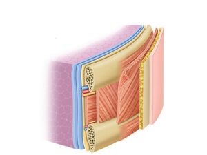
The thoracic wall layers
24,50 € Add to basketThoracic wall layers include skin, subcutaneous tissue,
intercostal muscles, and ribs. These structures protect
vital organs and allow chest movements required for
breathing.