Marrow
Showing 1–12 of 183 results
-
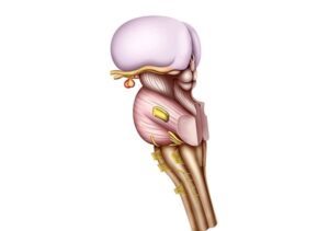
Anatomy Brainstem
24,50 € Add to basketThis illustration represents the anatomy of the
brainstem. First, it shows the inferior portion of
the brain, which connects the spinal cord with
higher structures.


-
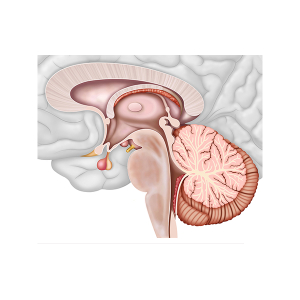
Anatomy of the brain
24,50 € Add to basketThe illustration displays the brainstem. First,
this structure corresponds to the lower portion
of the brain. Additionally, it connects the spinal
cord with the upper structures of the brain. -
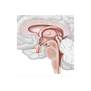
Anatomy of the brainstem
24,50 € Add to basketThe anatomy of the brainstem, also known as the brain
stem, is one of the most important structures of the central
nervous system. This region is located in the lower part of
the brain and establishes a direct connection between the
brain and the spinal cord..
-
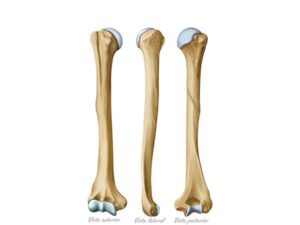
Anatomy of the humerus bone
24,50 € Add to basketAnatomy of the humerus, shown in three views. It extends from the
shoulder to the elbow. It consists of the head, neck, and shaft.
It articulates with the scapula at the shoulder and with the ulna and
radius at the elbow, allowing for a wide range of arm movements. -
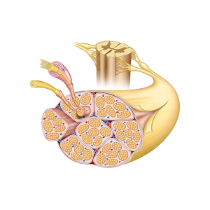
Anatomy of the nerve
24,50 € Add to basketThe anatomy of the nerve is composed of nerve fibers,
sheaths, and blood vessels. These fibers are made up
of axons and dendrites that transmit signals.
-
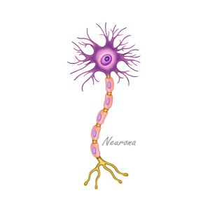
Anatomy of the neuron
24,50 € Add to basketThe anatomy of the neuron focuses on the structure
and function of these nerve cells. First, the neuron
consists of key parts such as the cell body, dendrites,
and axons.
-
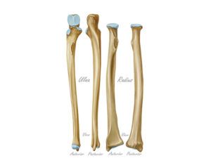
Anatomy of the radius and ulna bones
24,50 € Add to basketEs un hecho establecido hace demasiado tiempo que
un lector se distraerá con el contenido del texto de un<br />sitio mientras que mira su diseño. El punto de usar
Lorem Ipsum -
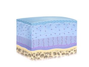
Articular cartilage structure
24,50 € Add to basketStructure of articular cartilage. It is a strong and elastic connective
tissue that covers the articular surfaces of bones in joints. It consists
of chondrocytes and extracellular matrix, providing cushioning and
facilitating smooth gliding of the joints. -
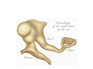
Auditory ossicles
24,50 € Add to basketThe ossicles of the middle ear. Also known as auditory ossicles,
these are three small bones in the middle ear: the malleus,
incus, and stapes. These bones transmit vibrations from the
eardrum to the inner ear, facilitating hearing. -
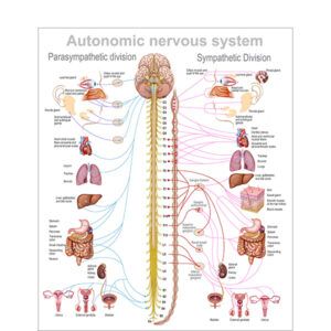
Autonomic nervous system
24,50 € Add to basketThe autonomic nervous system (ANS) is a vital regulatory
network responsible for controlling involuntary body functions.
Unlike other control pathways in the body, this system operates
automatically and does not require conscious effort..
-
Basal cerebral surface
24,50 € Add to basketThe basal cerebral surface refers to the lower part
of the brain, which rests on the base
of the skull. This illustration represents this
region from a lower perspective. -
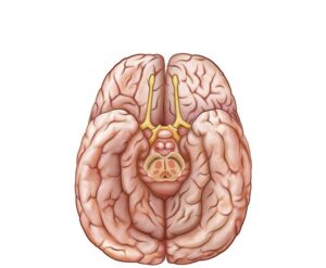
Basal surface of the brain
24,50 € Add to basketBasal surface of the brain. This illustration details the lower
part of the brain and the brainstem. The region coordinates
essential motor functions, sensory, and autonomic processes.
Its technical study is fundamental for the treatment of injuries
and various neurological disorders.