Medulla
Showing 1–12 of 65 results
-
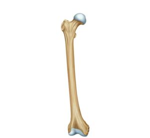
Anatomy of the femur bone
24,50 € Add to basketAnatomy of the femur bone. The longest and strongest bone in
the body is located between the hip and the knee. Its structure,
composed of head, neck, and shaft, facilitates body weight support
and musculoskeletal mobility through its connection with the pelvis
and the tibia.


-
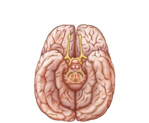
Basal surface of the brain
24,50 € Add to basketBasal surface of the brain. This illustration details the lower
part of the brain and the brainstem. The region coordinates
essential motor functions, sensory, and autonomic processes.
Its technical study is fundamental for the treatment of injuries
and various neurological disorders. -
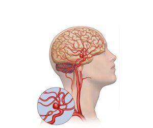
Cerebral arterial aneurysm
24,50 € Add to basketCerebral arterial aneurysm. Detailed medical illustration of a
human head, where weakened bulges in the artery are visualized.
This condition shows how vessels fill with blood, pressing on
surrounding tissue and creating a high risk of severe hemorrhage
in the patient. -
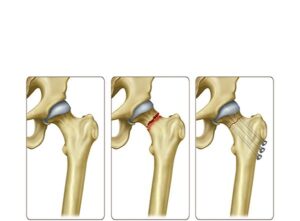
Femoral neck fracture treatment
24,50 € Add to basketFemoral neck fracture treatment involves internal fixation using
metal screws or nails to stabilize the bone and promote healing.
Moreover, it restores hip function. Treatment choice depends on
fracture severity and patient condition. -
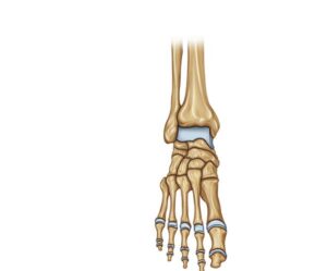
Frontal view of the foot
24,50 € Add to basketFrontal view of the foot. Technical representation of the bony
structures, including phalanges, metatarsals, and tarsal bones.
The image details the organization of the talus, calcaneus, and
cuneiforms, elements that provide the necessary anatomical base
for balance and locomotion. -
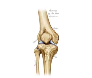
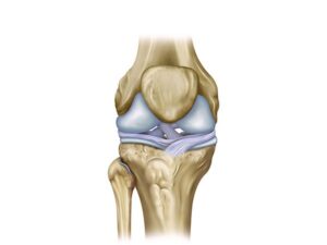
Frontal view of the knee
24,50 € Add to basketFrontal view of the knee. Technical illustration of the joint showing
the arrangement of the femur, tibia, fibula, and patella. The image
displays the articular surfaces and cartilage, key elements for flexion
and human locomotion in the activities of daily living. -
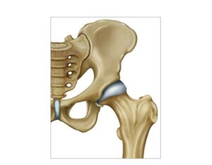
Hip and femur joint
24,50 € Add to basketHip and femur joint. The anatomical union between the acetabulum
and the femoral head allows the execution of flexion, extension, and
rotation movements. This bony structure supports the weight of the
torso and facilitates human locomotion, being vital for stability and
physical balance. -
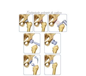
Hip osteoarthritis treatment
24,50 € Add to basketHip osteoarthritis and femoral neck treatment includes
physical therapy, medication, and prosthetic surgery. Moreover,
it aims to relieve pain and improve mobility. Treatment choice
depends on age, health status, and severity of joint damage. -
Knee cruciate ligament
24,50 € Add to basketKnee cruciate ligament. This anatomical structure joins the femur
with the tibia to provide internal stability. The anterior and posterior
cruciate ligaments are fundamental to preventing excessive bone
displacement, facilitating the diagnosis and treatment of common
joint injuries. -
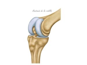
Knee function
24,50 € Add to basketKnee function. This joint acts as a complex hinge that facilitates
the flexion and extension of the leg. Its structure, composed of the
femur and tibia, supports body weight and guarantees the stability
necessary to walk, run, and perform daily activities safely. -
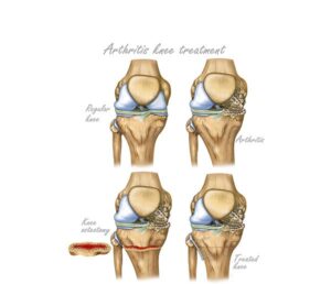
Knee osteotomy treatment
24,50 € Add to basketKnee osteotomy treatment is a surgical procedure that corrects
bone alignment to relieve pain and improve joint function. Moreover,
it redistributes load in early osteoarthritis and may delay the need for
knee replacement surgery. -
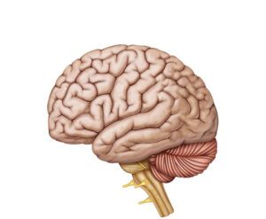
Lateral view of the brain
24,50 € Add to basketLateral view of the brain. This anatomical representation shows
the main sulci and lobes of the nervous system. The structure
includes the frontal, parietal, temporal, and occipital sectors. Each
area manages specific processes such as memory, sensory
perception, and cognition.