Medulla
Showing 13–24 of 65 results
-
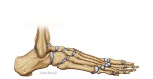
Lateral view of the foot
24,50 € Add to basketLateral view of the foot. Illustration of the bony anatomy showing
the connection of the tibia and fibula with the talus and calcaneus.
The image details the tarsal bones, such as the navicular and cuboid,
essential for maintaining balance, arch structure, and human mobility.


-
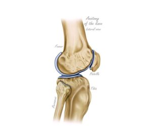
Lateral view of the knee
24,50 € Add to basketLateral view of the knee. This anatomical perspective details the
relationship between the femur, tibia, and patella. The illustration
highlights the articular surfaces and cartilages that allow for flexion
and extension of the leg, being fundamental for understanding
locomotion and skeletal stability. -
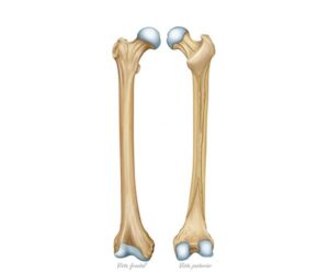
Morphology of the femur bone
24,50 € Add to basketMorphology of the femur bone. The longest bone in the thigh
presents a robust structure with a well-defined head, neck, and body.
Its frontal and posterior views reveal details such as the condyles and
the intertrochanteric crest, crucial elements for body support and
joint connection. -
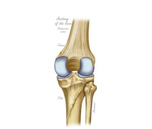
Posterior view of the knee
24,50 € Add to basketPosterior view of the knee. Technical illustration detailing the
back of the joint, including the femur, tibia, and fibula. The image
shows the structures necessary for flexion and leg biomechanics,
serving as an essential tool for evaluating functions and injuries. -
Rheumatoid arthritis
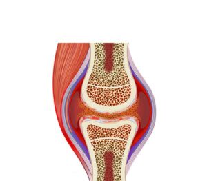
24,50 € Add to basketRheumatoid arthritis is an autoimmune disease causing chronic
inflammation and cartilage destruction. Moreover, it leads to pain,
stiffness, and joint deformity. Treatment includes disease-modifying
drugs, anti-inflammatory therapy, and physical rehabilitation. -
Subcapital femoral prosthesis
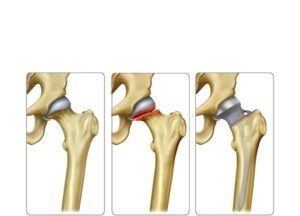
24,50 € Add to basketSubcapital femoral prosthesis is a surgical treatment for
severe fractures near the femoral head. Moreover, it replaces
damaged structures, relieving pain and restoring mobility.
Indication depends on patient age, health, and fracture severity. -
Synovial inflammation arthritis
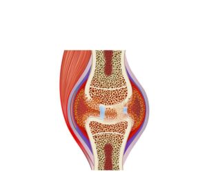
24,50 € Add to basketSynovial inflammation arthritis. This illustration shows the process
where the membrane lining the joints presents evident swelling.
The synovial tissue thickens due to rheumatoid arthritis or physical
wear, producing chronic pain symptoms and a significant loss of
joint mobility. -
Template
24,50 € Add to basketLorem Ipsum es simplemente el texto de relleno de las imprentas
y archivos de texto. Lorem Ipsum ha sido el texto de relleno estándar
de las industrias desde el año 1500, cuando un impresor (N. del T.
persona que se dedica a la imprenta) desconocido usó -
template (Copy)
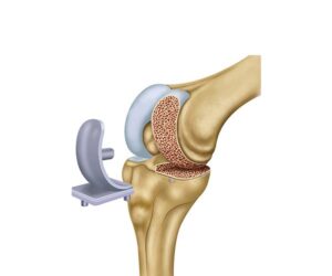
24,50 € Add to basketPartial knee replacement is a surgical procedure that replaces
only the damaged part of the knee joint. Moreover, it relieves pain
and improves mobility in localized osteoarthritis. Selection depends
on age, health status, and extent of joint damage. -
template (Copy)
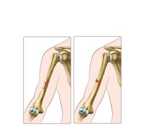
24,50 € Add to basketIncomplete and linear fracture includes partial or straight
bone breaks, commonly seen in trauma and pediatric patients.
Moreover, greenstick fractures occur due to bone flexibility and
may present with pain, swelling, and limited movement. -
The ankle pain
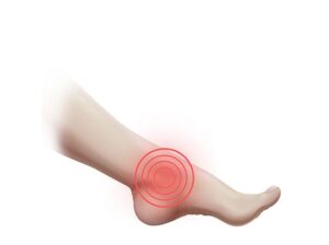
24,50 € Add to basketAnkle pain is a common condition caused by sprains, fractures,
or joint inflammation. Moreover, it can lead to swelling and difficulty
walking. Treatment includes rest, ice, physical therapy, and, in severe
cases, surgical intervention. -
The bone fracture and healing
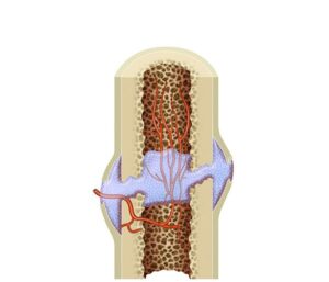
24,50 € Add to basketBone fracture and healing describe the repair process after
a bone break, including hematoma formation, callus development,
and remodeling. Moreover, it involves biological mechanisms that
restore bone structure and function.