Vision
Showing 25–36 of 67 results
-
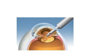
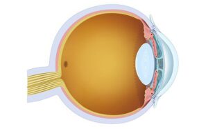
Lens extraction in cataracts
24,50 € Add to basketLens extraction in cataracts is a surgical procedure used
in advanced cases. This representation shows the cornea,
lens, aqueous humor, retina, and optic nerve.


-
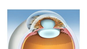
Lens of the eye
24,50 € Add to basketThe crystalline lens is a transparent and flexible structure
located behind the iris that focuses light onto the retina.
Through accommodation, controlled by the ciliary muscles,
the lens changes shape to focus on objects at different
distances. -
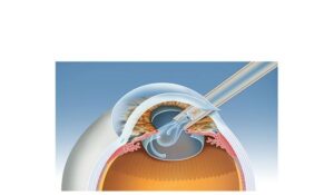
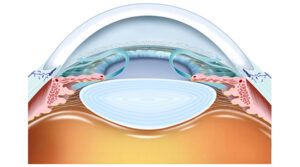
Lens replacement in cataracts
24,50 € Add to basketThe lens replacement in cataract surgery illustrates the final stage,
where the cloudy lens is replaced with an artificial intraocular lens.
After removing the lens using phacoemulsification, the new lens is
implanted through a small incision in the cornea. -
Macula and fovea
24,50 € Add to basketThe macula and fovea are central retinal regions responsible
for detailed vision. The macula supports high visual acuity,
while the fovea contains the highest density of cone cells,
allowing precise color perception and sharp central vision. -
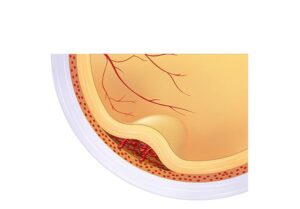
Macular Degeneration
24,50 € Add to basketMacular Degeneration (AMD) illustrates how the condition affects
the macula, the central part of the retina responsible for detailed
vision. Aging can damage the cells of the macula, leading to blurred
vision or loss of central vision. -
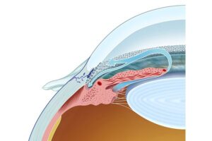
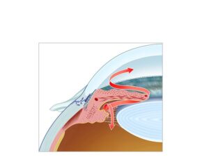
Normal aqueous drainage
24,50 € Add to basketNormal aqueous humor drainage describes the flow from
the ciliary bodies through the trabecular meshwork and
Schlemm’s canal regulates eye pressure. -
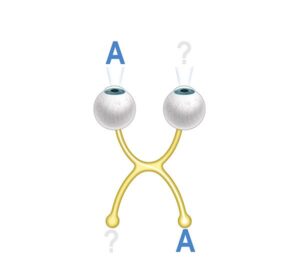
Ocular amblyopia
24,50 € Add to basketOcular amblyopia, also known as “lazy eye“, is a condition in
which the eye does not develop properly during childhood. May
result in reduced vision in that eye, even with optical correction. -
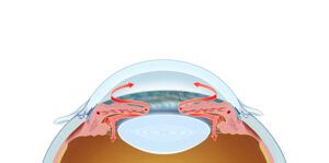
Ocular aqueous drainage
24,50 € Add to basketAqueous humor drainage describes the flow of fluid
from the ciliary body through the trabecular meshwork
and the canal of Schlemm to regulate eye pressure. -
Ocular drainage in glaucoma
24,50 € Add to basketOcular drainage in glaucoma shows how obstruction or dysfunction
in the eye’s drainage systems can lead to an increase in intraocular
pressure. In a healthy eye, aqueous humor flows properly through the
trabecular meshwork and Schlemm’s canal. -
Ocular drainage obstruction
24,50 € Add to basketThe blockage of the eye’s drainage prevents the proper flow of
aqueous humor, which can increase intraocular pressure. In a
healthy eye, aqueous humor flows normally through the drainage
channels, such as the trabecular meshwork and Schlemm’s canal. -
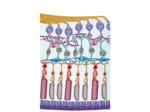
Photoreceptors cones and rods
24,50 € Add to basketPhotoreceptors in the retina include cones and rods. Cones
enable detailed color vision in bright light, while rods provide
sensitivity to dim light and support peripheral vision. -
Physiology of aqueous humor
24,50 € Add to basketAqueous humor physiology explains how this fluid nourishes
the cornea and lens while maintaining intraocular pressure
through circulation and drainage.