Vision
Showing 1–12 of 67 results
-
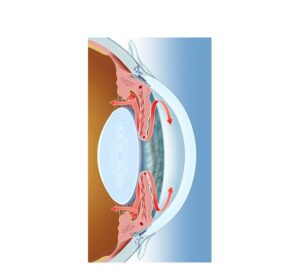
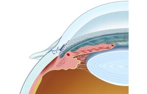
Altered aqueous humor drainage
24,50 € Add to basketAlteration of aqueous humor drainage in an eye with glaucoma. It
shows how a blockage in the trabecular meshwork or in Schlemm’s
canal prevents proper fluid flow, causing an increase in intraocular
pressure.


-
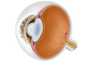
Anatomy of the eye
24,50 € Add to basketEye anatomy includes the cornea, iris, pupil, lens, and retina
and the optic nerve that work together to capture light and create
visual perception. -
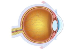
Anatomy of the eye
24,50 € Add to basketHuman eye anatomy includes structures such as the cornea,
iris, pupil, lens, retina, and optic nerve. These components work
together to focus light, convert it into nerve signals, and allow
the brain to perceive visual images clearly. -
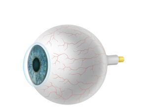
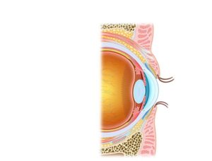
Anatomy of the eyeball
24,50 € Add to basketThe eyeball is a spherical structure composed of layers
such as the sclera, cornea, and retina. These structures
allow light to enter the eye, focus images, and convert
light into nerve signals for vision. -
Anatomy of the eyelids
24,50 € Add to basketThe eyelids protect the eye and help maintain ocular
moisture through blinking. Eyelashes and sebaceous
glands prevent particles from entering the eye and
support tear film stability. -
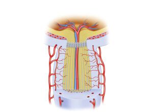
Anatomy of the Optic Nerve
24,50 € Add to basketThe optic nerve connects the retina to the brain and
transmits electrical signals generated by photoreceptors.
These signals are processed in the visual cortex, allowing
the brain to interpret visual images. -
Aqueous humor of the eye
24,50 € Add to basketAqueous humor is a clear fluid that nourishes the <br />cornea and lens while maintaining intraocular pressure
and eye shape.</p> -
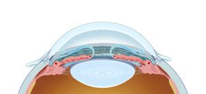
Aqueous humour drainage
24,50 € Add to basketAqueous humor drainage describes the flow of fluid from
the ciliary bodies through the trabecular meshwork and
Schlemm’s canal to the venous system. -
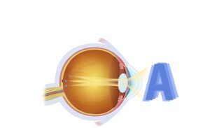
Astigmatism vision
24,50 € Add to basketAstigmatism affects vision. People with astigmatism see things
blurry and distorted, and have trouble focusing both up close and
far away. Glasses, contact lenses, and refractive procedures like
laser surgery can correct astigmatism. -
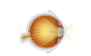
Cataract focusing
24,50 € Add to basketThe cataract focuses. This is affected when the lens, the eye’s
natural lens, becomes cloudy. Cataracts can blur vision, cause hazy
sight, and impair the ability to focus properly. -
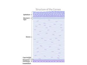
Cornea and ocular surface
24,50 € Add to basketThe cornea and ocular surface allow light to enter the eye
and contribute to focusing images on the retina. In addition,
the cornea protects the eye and maintains visual clarity
through its transparency and curved structure. -
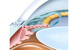
Detail of Schlemm’s canal
24,50 € Add to basketSchlemm’s canal collects aqueous humor filtered by the
trabecular meshwork and directs it to the venous system,
helping regulate eye pressure.