Anatomy
Showing 349–360 of 678 results
-
Neuromuscular synapse myasthenia
24,50 € Add to basketNeuromuscular synapse myasthenia explains muscle weakness
caused by antibodies blocking acetylcholine. This disruption in the
neuromuscular junction generates fatigue and failures in nerve
communication that affect voluntary movement.


-
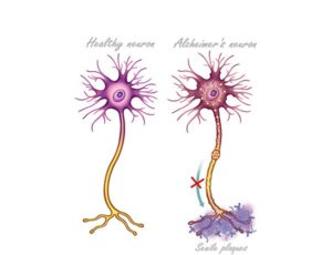
Neuron in Alzheimer’s disease
24,50 € Add to basketThe neuron in Alzheimer’s disease has an altered function in
information transmission. Moreover, under normal conditions,
neurons communicate efficiently. Additionally, they support
learning and memory. -
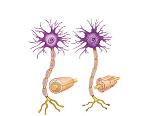
Neuron with demyelinating disease
24,50 € Add to basketThe neuron with demyelinating disease has an altered function in
nervous system transmission. Moreover, under normal conditions,
myelin acts as an insulator. Additionally, it allows rapid conduction
of nerve impulses. It ensures efficient neuronal communication. -
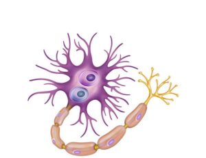
Neuron with Lewy bodies
24,50 € Add to basketThe neuron with Lewy bodies has an altered function in neuronal
communication. Moreover, under normal conditions, neurons
transmit signals efficiently. Additionally, they maintain cellular
integrity. Furthermore, they support proper nervous system function. -
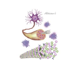
Neuronal alteration in Alzheimer’s disease
24,50 € Add to basketThe neuronal alteration in Alzheimer’s disease has an altered function
in the nervous system. Moreover, under normal conditions, neurons
communicate efficiently. Additionally, they support memory consolidation
and learning. -
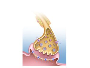
Neuronal synapse
24,50 € Add to basketNeuronal synapse is the connection between neurons
where neurotransmitters transmit signals across the
synaptic cleft, enabling nervous system communication. -
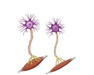
Neuronal synapse in ALS
24,50 € Add to basketThe neuronal synapse in ALS has an altered function in nervous
system communication. Moreover, under normal conditions,
neurons transmit electrical impulses efficiently. Additionally, they
enable muscle activation. -
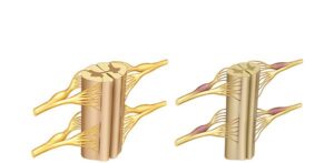
Normal and ataxic spinal cord
24,50 € Add to basketThe normal and ataxic spinal cord has an altered function in motor
coordination. Moreover, under normal conditions, the spinal cord
transmits signals efficiently. Additionally, it coordinates communication
between the brain and muscles. -
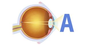
Normal eye focus
24,50 € Add to basketNormal eye focus. This illustration presents the path of light
through the cornea and lens to the retina. The graphic shows the
transformation of light signals into electrical impulses for brain
processing, ensuring a sharp and detailed vision of the
surroundings. -
Nucleosome structure
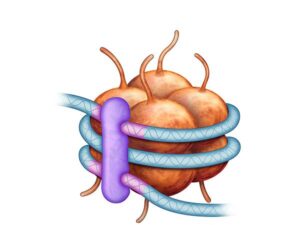
24,50 € Add to basketThe nucleosome is the basic unit of chromatin, formed by
DNA wrapped around histones, enabling DNA compaction,
protection, and gene regulation. -
Ocular amblyopia
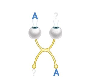
24,50 € Add to basketOcular amblyopia, also known as “lazy eye“, is a condition in
which the eye does not develop properly during childhood. May
result in reduced vision in that eye, even with optical correction. -
Ocular drainage in glaucoma
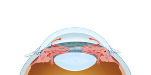
24,50 € Add to basketOcular drainage in glaucoma shows how obstruction or dysfunction
in the eye’s drainage systems can lead to an increase in intraocular
pressure. In a healthy eye, aqueous humor flows properly through the
trabecular meshwork and Schlemm’s canal.