Lungs
Showing 1–12 of 109 results
-
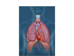
A cardiopulmonary anatomical illustration
24,50 € Add to basketA cardiopulmonary anatomical illustration shows how
the heart and lungs work together to circulate oxygenated
blood, remove carbon dioxide, and maintain essential
body functions.


-
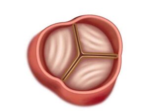
A closed aortic valve
24,50 € Add to basketA closed aortic valve prevents blood from flowing
backward from the aorta into the left ventricle during
diastole, ensuring proper one-way blood circulation
through the cardiovascular system. -
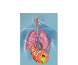
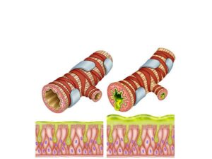
Acute bronchitis
24,50 € Add to basketAcute bronchitis. Inflammation of the bronchi generally
caused by viral infections, which produce cough, mucus, and
respiratory difficulty, being a common condition after
colds or flu. -
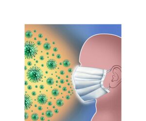
Airborne transmission
24,50 € Add to basketAirborne transmission occurs when infectious particles are
transmitted through the air. Preventive measures are essential.
to reduce its spread. The use of masks protects and reduces
the spread of respiratory infections. -
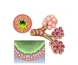
Altered bronchial mucosa
24,50 € Add to basketAltered bronchial mucosa. It represents the increase in secretion
of mucus by goblet cells and bronchial inflammation, causing
airflow obstruction, persistent cough, and respiratory difficulty,
typical in bronchitis. -
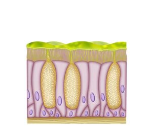
Altered mucous cells
24,50 € Add to basketAltered mucous cells. They represent the hyperactivity of the
goblet cells with excessive production of thick mucus, which
which causes obstruction of the airways, persistent cough
and respiratory difficulty in bronchitis. -
Alveolar mucus
24,50 € Add to basketAlveolar mucus. It represents the excessive and thick accumulation
of mucus in the respiratory epithelium, caused by inflammation, which
hinders airflow and causes persistent cough and respiratory problems. -
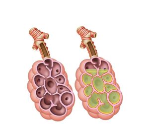
Alveolar pneumonia infection
24,50 € Add to basketInfectious alveolar pneumonia is an inflammation of the alveoli.
caused by microorganisms. It causes fluid accumulation, hindering
gas exchange and generating respiratory symptoms. -
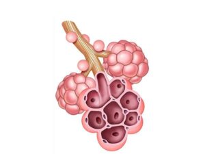
Alveolar structure
24,50 € Add to basketThe alveolar structure consists of small air sacs called
alveoli located in the lungs. These microscopic structures
allow oxygen to enter the bloodstream and carbon dioxide
to leave the body. -
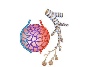
Alveoli and bronchioles
24,50 € Add to basketAlveoli and bronchioles are lung structures where
gas exchange occurs. They allow oxygen to enter the
blood and carbon dioxide to leave the body during
breathing. -
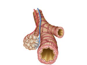
Anatomy of bronchioles and alveoli
24,50 € Add to basketBronchioles and alveoli are small respiratory structures
within the lungs. Bronchioles lack hyaline cartilage and
are surrounded by smooth muscle, while alveoli are tiny
air sacs where oxygen and carbon dioxide exchange occurs. -
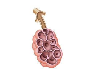
Anatomy of the alveolus
24,50 € Add to basketThe anatomy of the alveolus shows an air-filled cavity
with walls made of simple squamous epithelium. Elastic
and collagen fibers support pulmonary capillaries and
enable gas exchange.