Sight
Showing 13–24 of 35 results
-
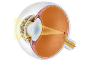
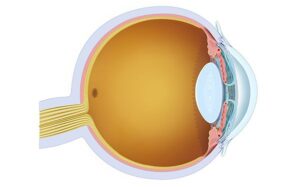
Eye with normal vision.
24,50 € Add to basketNormal vision occurs when light enters the eye through
the cornea, passes through the pupil, and is focused by the
lens onto the retina. Through accommodation, the eye can
adjust focus for near and distant objects, allowing clear
and sharp visual perception.


-
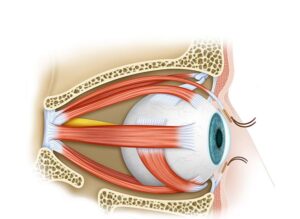
Human extraocular muscles
24,50 € Add to basketHuman extraocular muscles control the movement of the eyeball.
These six muscles work together to produce vertical, horizontal,
and rotational eye movements, allowing both eyes to coordinate
their position and maintain accurate visual tracking. -
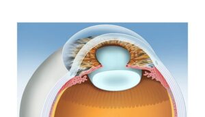
Lens of the eye
24,50 € Add to basketThe crystalline lens is a transparent and flexible structure
located behind the iris that focuses light onto the retina.
Through accommodation, controlled by the ciliary muscles,
the lens changes shape to focus on objects at different
distances. -
Macula and fovea
24,50 € Add to basketThe macula and fovea are central retinal regions responsible
for detailed vision. The macula supports high visual acuity,
while the fovea contains the highest density of cone cells,
allowing precise color perception and sharp central vision. -
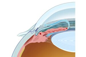
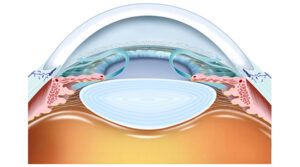
Normal aqueous drainage
24,50 € Add to basketNormal aqueous humor drainage describes the flow from
the ciliary bodies through the trabecular meshwork and
Schlemm’s canal regulates eye pressure. -
Ocular aqueous drainage
24,50 € Add to basketAqueous humor drainage describes the flow of fluid
from the ciliary body through the trabecular meshwork
and the canal of Schlemm to regulate eye pressure. -
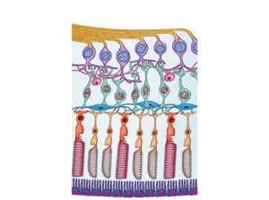
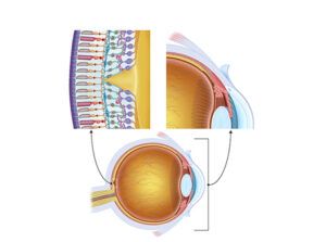
Photoreceptors cones and rods
24,50 € Add to basketPhotoreceptors in the retina include cones and rods. Cones
enable detailed color vision in bright light, while rods provide
sensitivity to dim light and support peripheral vision. -
Physiology of aqueous humor
24,50 € Add to basketAqueous humor physiology explains how this fluid nourishes
the cornea and lens while maintaining intraocular pressure
through circulation and drainage. -
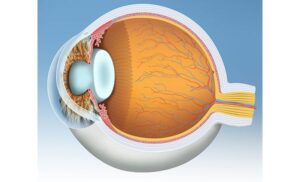
Physiology of the eye
24,50 € Add to basketEye physiology explains how the cornea, iris, pupil,
lens, retina, and optic nerve work together to capture
light and create vision. -
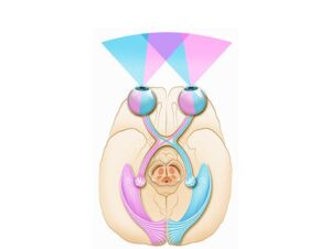
Physiology of the visual system
24,50 € Add to basketThe physiology of the visual system explains how the eye, The
optic nerve and the brain work together to convert light into
nerve signals that allow humans to perceive images and
interpret the visual world. -
Physiology of vision
24,50 € Add to basketVisual physiology explains how the eye captures light and
converts it into nerve signals processed by the brain. This
process includes focusing light on the retina, transmitting
signals through the optic nerve and perceiving color, contrast
and brightness. -
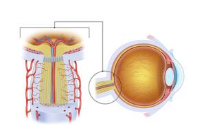
Physiology Optic Nerve
24,50 € Add to basketThe optic nerve carries visual signals from the retina to
the brain. These electrical impulses are processed in the
visual cortex, allowing the brain to interpret light, color,
and shapes as visual images.