Sight
Showing 25–35 of 35 results
-
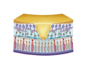
Retina and macula structure
24,50 € Add to basketThe retina and macula contain photoreceptor cells that
detect light and enable vision. The macula and its central
region, the fovea, provide detailed central vision and
accurate color perception.


-
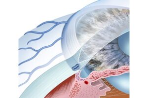
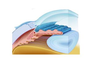
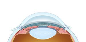
Schlemm’s canal
24,50 € Add to basketThe canal of Schlemm drains aqueous humor from the
trabecular meshwork into the venous system, helping
regulate intraocular pressure. -
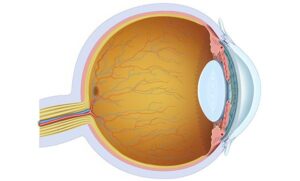
Structure of the eye
24,50 € Add to basketThe structure of the eye includes the cornea, iris, pupil,
lens, retina, and optic nerve that capture light and transmit
visual information. -
The cornea and iris
24,50 € Add to basketThe cornea and iris are anterior eye structures that control
light entry and help focus images on the retina. The cornea
refracts light while the iris regulates pupil size to adapt vision
to different lighting conditions. -
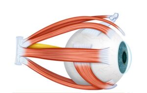
The eye muscles
24,50 € Add to basketOcular muscles control eye position and movement. Six
muscles work together to produce vertical, horizontal, and
rotational movements, allowing coordinated gaze and stable
visual tracking. -
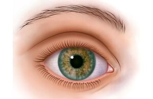
The human eye
24,50 € Add to basketThe external anatomy of the eye includes the cornea,
sclera, iris, pupil, and eyelids that protect the eye and
regulate the entry of light. -
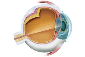
The ocular anatomy
24,50 € Add to basketOcular anatomy includes the cornea, iris, pupil, lens,
retina, and optic nerve. These structures work together
to focus light, convert it into nerve signals, and allow
visual perception. -
The physiology of vision
24,50 € Add to basketVisual physiology explains how the eye focuses light on
the retina converts it into nerve signals that the brain
interprets as images, allowing the perception of color,
shape and detail. -
The Schlemm’s Canal
24,50 € Add to basketThe canal of Schlemm drains aqueous humor from the
trabecular meshwork into the venous system, helping
regulate intraocular pressure. -
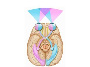
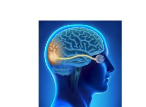
The visual system
24,50 € Add to basketThe visual system includes the eye, optic nerve, and
visual cortex. These structures convert light into nerve
signals that allow the brain to perceive images, colors,
shapes, and movement. -
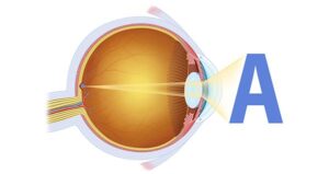
Visualization of the object
24,50 € Add to basketObject visualization occurs when light enters the eye,
is focused on the retina and converted into nerve signals
that travel through the optic nerve to the brain, allowing
the perception of colors, shapes, and fine visual details.