Anatomy
Showing 121–132 of 678 results
-
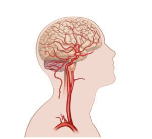
Cerebral blood circulation
24,50 € Add to basketCerebral blood circulation. This illustration represents the flow
of oxygenated blood from the arteries to the capillaries for nutrient
exchange. The graphic shows how veins return deoxygenated blood
to the heart, facilitating the understanding of cellular perfusion.


-
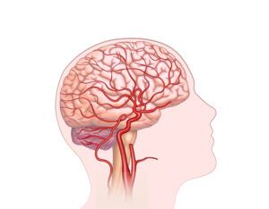
Cerebral blood flow
24,50 € Add to basketLorem Ipsum es simplemente el texto de relleno de las imprentas
y archivos de texto. Lorem Ipsum ha sido el texto de relleno estándar
de las industrias desde el año 1500, cuando un impresor (N. del T.
persona que se dedica a la imprenta) desconocido usó -
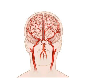
Cerebral carotid irrigation
24,50 € Add to basketCerebral carotid irrigation. This illustration shows the blood
supply through the internal carotids and vertebral arteries connected
by the Circle of Willis. The diagram details how the internal carotid
nourishes the forebrain and the upper lobes for cognitive function. -
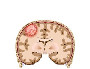
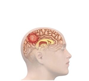
Cerebral meningioma tumor
24,50 € Add to basketCerebral meningioma tumor. This realistic illustration details
a tumor originating in the meningeal membranes and its impact on
brain structure. The content highlights the importance of this visual
resource for clinical diagnosis, surgical planning, and education. -
Cerebral meningioma tumor
24,50 € Add to basketCerebral meningioma tumor. This realistic illustration details
a tumor originating in the meningeal membranes and its impact on
brain structure. The content highlights the importance of this visual
resource for clinical diagnosis, surgical planning, and education. -
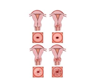
Cervical cancer stages
24,50 € Add to basketCervical cancer stages describe tumor progression from localized
abnormal cells to spread into nearby and distant organs. This
classification is essential for guiding treatment and determining
prognosis. -
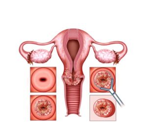
Cervical tumor treatment
24,50 € Add to basketCervical tumor treatment. This medical procedure focuses on
the elimination of cancerous cells in the cervix through surgery or
chemotherapy. The choice of therapy depends on the stage of the
disease. The process guarantees professional management to
restore health. -
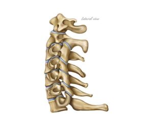
Cervical vertebrae lateral view
24,50 € Add to basketThe lateral views of the cervical vertebrae highlight the seven
bony structures in the neck region, numbered C1 through C7. The
natural curves of the spine and anatomical features such as the
vertebral bodies, neural arches, and spinous processes are visible. -
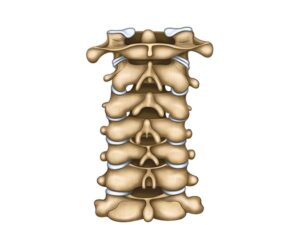
Cervical vertebrae posterior view
24,50 € Add to basketPosterior view of the cervical vertebrae. These are the
bony structures in the neck region of the spine. They are
located at the back of the neck and consist of seven
vertebrae numbered C1 through C7. -
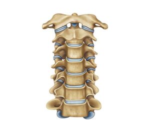
Cervical vertebrae, frontal view
24,50 € Add to basketCervical vertebrae in frontal view. The seven vertebrae that
make up the cervical region of the spine are shown. Numbered
C1 through C7, these bony structures form the neck, providing
support and flexibility. -
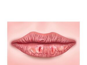
Chapped lips in Sjögren
24,50 € Add to basketChapped lips in Sjögren. This illustration details the dryness and
labial fissures caused by xerostomia in patients with this autoimmune
pathology. The content describes how the lack of saliva damages tissues
and highlights the importance of constant hydration and emollients. -
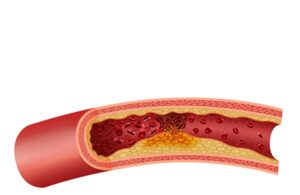
Cholesterol plaque rupture
24,50 € Add to basketRupture of cholesterol plaque. The atherosclerotic plaque ruptures
and generates a clot that blocks blood flow, increasing the risk of heart
attack and stroke, requiring urgent medical attention.