Neuron
Showing 109–120 of 129 results
-
Template (Copy)
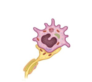
24,50 € Add to basketMerkel cells are specialized skin mechanoreceptors located
in the epidermis, associated with nerve endings, enabling
detection of touch, pressure, and texture.


-
Template (Copy)
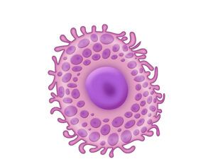
24,50 € Add to basketThe mast cell is an immune cell that releases histamine and
mediators, playing a key role in allergic reactions, inflammation,
and defense against pathogens. -
The basal ganglia
24,50 € Add to basketThe basal ganglia are a set of brain nuclei
that play a crucial role in regulating movement
and motor coordination. They are located on
either side of the thalamus. -
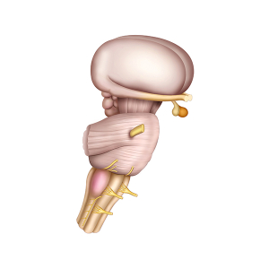
The brain stem
24,50 € Add to basketThis illustration shows the brainstem. First, this
structure is vital for the basic functions of the
nervous system. Additionally, it is located at the
base of the brain and connects the encephalon
with the spinal cord. -
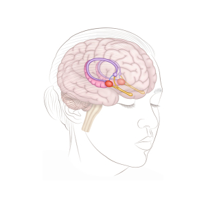
The brain stem
24,50 € Add to basketRepresentation of the human head and the limbic
system shows the location of these structures
within the brain and key components such as the
hippocampus, amygdala, and hypothalamus. -
The brain tonsils
24,50 € Add to basketThe brain tonsils. This representation of the human
head shows the location and function of these key
structures in the brain. First, the amygdalae are
located in the temporal lobe. -
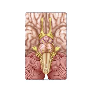
The brainstem
24,50 € Add to basketThe brainstem provides a detailed view of the
structures in the lower part of the encephalon.
First, it regulates vital functions such as breathing
and heart rate.
-
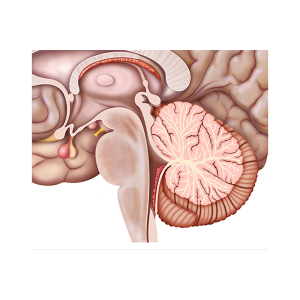
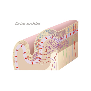
The cerebellar cortex
24,50 € Add to basketThis illustration shows the cerebellar cortex, a
structure essential for motor coordination. First,
it is important to note that it is organized into
three well-defined layers. -
The cerebral amygdala
24,50 € Add to basketThe amygdala is a brain structure located in the
temporal lobe. First, it is essential for processing
emotions, especially fear and aggression. -
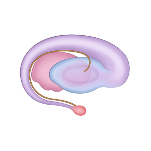
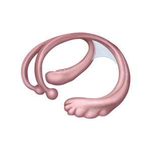
The cerebral fornix
24,50 € Add to basketThe structure of the cerebral fornix constitutes
a pathway of nerve fibers that connects various
regions of the brain. Involved in areas of
memory, emotions, and communication. -
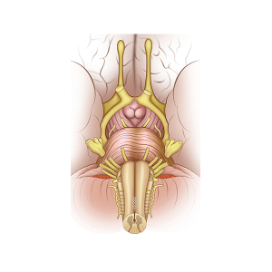
The cranial nerves
24,50 € Add to basketThe cranial nerves are a key part of the nervous system.
First, they connect the brain to the spinal cord and
control vital functions. Additionally, they contain
nuclei that regulate sensory and motor functions
of the head and neck. -
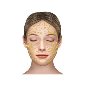
The facial nerves
24,50 € Add to basketThe facial nerves are shown on a female face, where
their distribution and function are clearly highlighted.
First, they correspond to cranial nerve VII.