Skeleton
Showing 1–12 of 134 results
-
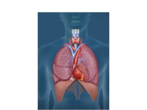
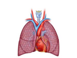
A cardiopulmonary anatomical illustration
24,50 € Add to basketA cardiopulmonary anatomical illustration shows how
the heart and lungs work together to circulate oxygenated
blood, remove carbon dioxide, and maintain essential
body functions.


-
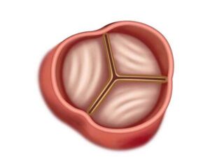
A closed aortic valve
24,50 € Add to basketA closed aortic valve prevents blood from flowing
backward from the aorta into the left ventricle during
diastole, ensuring proper one-way blood circulation
through the cardiovascular system. -
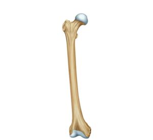
Anatomy of the femur bone
24,50 € Add to basketAnatomy of the femur bone. The longest and strongest bone in
the body is located between the hip and the knee. Its structure,
composed of head, neck, and shaft, facilitates body weight support
and musculoskeletal mobility through its connection with the pelvis
and the tibia. -
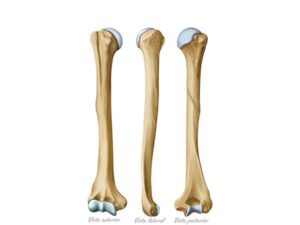
Anatomy of the humerus bone
24,50 € Add to basketAnatomy of the humerus, shown in three views. It extends from the
shoulder to the elbow. It consists of the head, neck, and shaft.
It articulates with the scapula at the shoulder and with the ulna and
radius at the elbow, allowing for a wide range of arm movements. -
Anatomy of the Pericardium
24,50 € Add to basketThe anatomy of the pericardium shows the layers that
surround the heart. It includes the fibrous pericardium
and the serous pericardium, divided into parietal and
visceral layers that protect the heart and allow smooth
cardiac movement. -
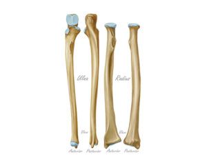
Anatomy of the radius and ulna bones
24,50 € Add to basketEs un hecho establecido hace demasiado tiempo que
un lector se distraerá con el contenido del texto de un<br />sitio mientras que mira su diseño. El punto de usar
Lorem Ipsum -
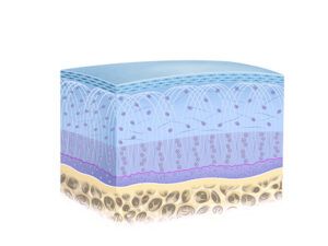
Articular cartilage structure
24,50 € Add to basketStructure of articular cartilage. It is a strong and elastic connective
tissue that covers the articular surfaces of bones in joints. It consists
of chondrocytes and extracellular matrix, providing cushioning and
facilitating smooth gliding of the joints. -
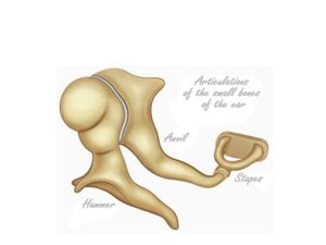
Auditory ossicles
24,50 € Add to basketThe ossicles of the middle ear. Also known as auditory ossicles,
these are three small bones in the middle ear: the malleus,
incus, and stapes. These bones transmit vibrations from the
eardrum to the inner ear, facilitating hearing. -
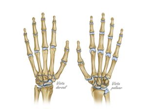
Bone anatomy of the hand
24,50 € Add to basketBone anatomy of the hand. This image shows both the palm and
the back of the hand. It illustrates the complex bone structure,
which includes the metacarpals, phalanges, and carpal bones. The
metacarpals form the base of the fingers, while the phalanges
make up the fingers themselves. -
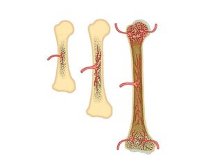
Bone growth and development
24,50 € Add to basketBone growth. This dynamic, regulated process in which bones
increase in length and thickness. In the growth zones, known as
growth plates or epiphyses, cartilage cells divide and transform
into bone tissue. -
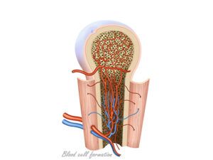
Bone marrow hematopoiesis
24,50 € Add to basketBone marrow hematopoiesis. The vital process of blood cell
formation in the bone marrow. This soft tissue, located inside
the bones, produces stem cells that differentiate into red blood
cells, white blood cells, and platelets. -
Cardiopulmonary anatomy
24,50 € Add to basketCardiopulmonary anatomy describes how the heart,
lungs, and blood vessels work together to circulate
oxygen, remove carbon dioxide, and support cellular
metabolism and physiological balance.