Skeleton
Showing 25–36 of 134 results
-
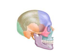
Facial and cranial bones
24,50 € Add to basketThe facial and cranial bones form the bony structure of
the head, protecting the brain and providing support for the
facial muscles. The skull is composed of bones such as the
frontal, parietal, temporal, and occipital bones.


-
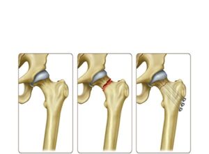
Femoral neck fracture treatment
24,50 € Add to basketFemoral neck fracture treatment involves internal fixation using
metal screws or nails to stabilize the bone and promote healing.
Moreover, it restores hip function. Treatment choice depends on
fracture severity and patient condition. -
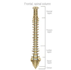
Frontal organization of the spine
24,50 € Add to basketThe frontal vertebral column highlights its crucial role as the
foundation for the mobility and stability of the human body.
It is composed of cervical (C1-C7), thoracic (T1-T12), and lumbar
(L1-L5), and sacral (S1-S5) vertebrae, as well as the coccyx. -
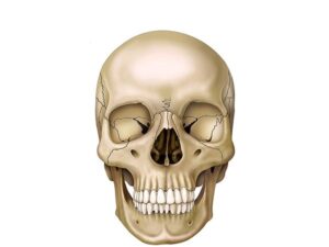
Frontal skull view
24,50 € Add to basketFrontal View of the Skull. In this view, the skull reveals the complexity
of its bony structure, which forms the facial anatomy and protects the
brain. Cranial bones such as the frontal and maxillary bones define
the facial appearance. -
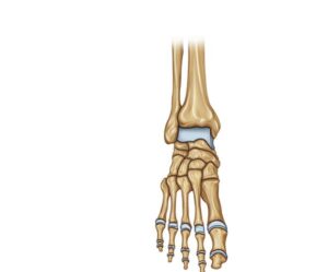
Frontal view of the foot
24,50 € Add to basketFrontal view of the foot. Technical representation of the bony
structures, including phalanges, metatarsals, and tarsal bones.
The image details the organization of the talus, calcaneus, and
cuneiforms, elements that provide the necessary anatomical base
for balance and locomotion. -
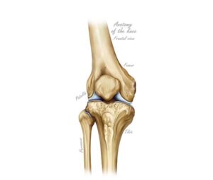
Frontal view of the knee
24,50 € Add to basketFrontal view of the knee. Technical illustration of the joint showing
the arrangement of the femur, tibia, fibula, and patella. The image
displays the articular surfaces and cartilage, key elements for flexion
and human locomotion in the activities of daily living. -
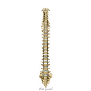
Frontal view of the spine
24,50 € Add to basketThe spinal column. It highlights its importance as a fundamental
pillar for the body’s mobility and stability. It consists of the cervical
vertebrae (C1–C7), thoracic vertebrae (T1–T12), lumbar vertebrae
(L1–L5), sacral vertebrae (S1–S5), and the coccyx. -
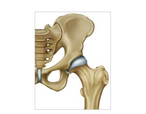
Hip and femur joint
24,50 € Add to basketHip and femur joint. The anatomical union between the acetabulum
and the femoral head allows the execution of flexion, extension, and
rotation movements. This bony structure supports the weight of the
torso and facilitates human locomotion, being vital for stability and
physical balance. -
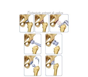
Hip osteoarthritis treatment
24,50 € Add to basketHip osteoarthritis and femoral neck treatment includes
physical therapy, medication, and prosthetic surgery. Moreover,
it aims to relieve pain and improve mobility. Treatment choice
depends on age, health status, and severity of joint damage. -
Human knee diagram
24,50 € Add to basketThe human knee. It displays its complex anatomical structure,
composed of bones, ligaments, cartilage, and muscles. The knee is
a vital joint that enables movements such as flexion and extension. -
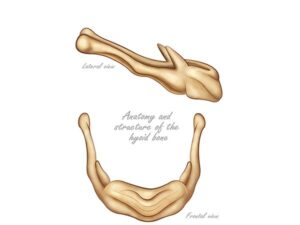
Hyoid bone
24,50 € Add to basketThe hyoid bone. It is a U-shaped structure located at the base
of the neck that does not articulate with other bones. It is situated
between the jaw and the larynx and is essential for functions such
as swallowing and speech. -
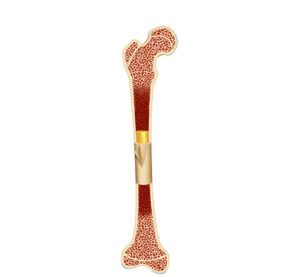
Internal anatomy of the femur
24,50 € Add to basketInternal structure of the femur. It highlights anatomical features such
as the bone marrow, the femoral neck, the bone trabeculae, and the
proximal and distal epiphyses. This diagram provides an overview of
the internal structure of the femur, with biomechanics and blood cells
production playing a fundamental role.