Skeleton
Showing 61–72 of 134 results
-
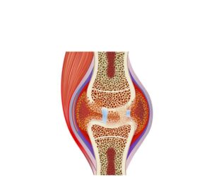
Rheumatoid arthritis
24,50 € Add to basketRheumatoid arthritis is an autoimmune disease causing chronic
inflammation and cartilage destruction. Moreover, it leads to pain,
stiffness, and joint deformity. Treatment includes disease-modifying
drugs, anti-inflammatory therapy, and physical rehabilitation.


-
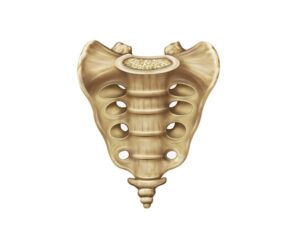
Sacrum frontal view
24,50 € Add to basketThe sacrum, viewed from the front, presents the fused triangular
structure at the base of the spine. Numbered S1-S5, the sacrum
forms the sacroiliac joint with the pelvis; the sacral crests and sacral
foramina are visible. -
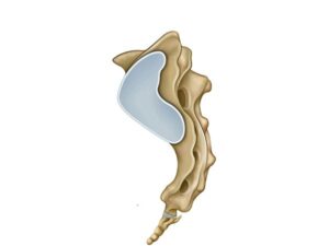
Sacrum lateral view
24,50 € Add to basketLateral view of the sacrum, with a triangular and fused structure
located at the base of the spine. Numbered S1-S5, the sacrum connects
to the pelvis and forms the sacroiliac joint. Lateral view of the sacrum.
The sacral crests and sacral foramina are visible. -
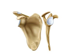
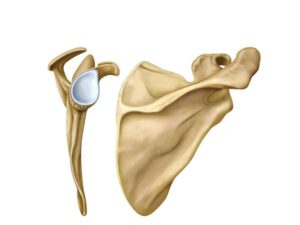
Scapula frontal view
24,50 € Add to basketFrontal view of the scapula. Also known as the shoulder blade,
this illustration shows this broad, triangular bone located on the
back. Notable features include the scapular spine, the glenoid
cavity, and the acromion. -
Scapula posterior view
24,50 € Add to basketPosterior view of the scapula. This illustration shows
this broad, triangular bone is located on the back.
Also known as the scapula, it features the scapular spine,
the glenoid cavity, and the acromion. -
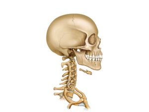
Skull and cervical vertebrae
24,50 € Add to basketThe skull and cervical vertebrae. The skull, composed of
the frontal, parietal, and occipital bones, protects the brain and
houses the sensory organs. The cervical vertebrae, located in the
neck region, form the spinal column. -
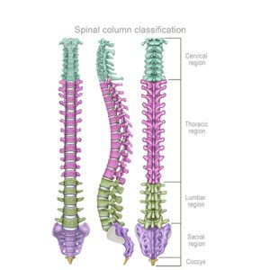
Spinal column classification
24,50 € Add to basketThe spine is classified based on its anatomical segments:
cervical, thoracic, lumbar, sacral, and coccygeal. The cervical
region (7 vertebrae, C1–C7), the thoracic region (12 vertebrae,
T1–T12), the lumbar region (5 vertebrae, L1–L5), and the sacral
region (5 vertebrae, S1–S5). -
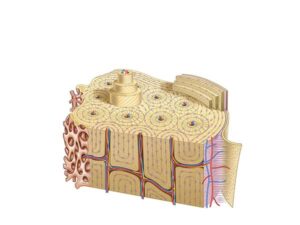
Spongy bone structure
24,50 € Add to basketStructure of cancellous bone. Also known as trabecular bone
tissue, it forms the inner part of bones and is characterized by a
porous, network-like structure. It consists of bone trabeculae
arranged irregularly, creating spaces filled with bone marrow. -
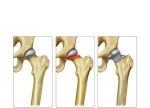
Subcapital femoral prosthesis
24,50 € Add to basketSubcapital femoral prosthesis is a surgical treatment for
severe fractures near the femoral head. Moreover, it replaces
damaged structures, relieving pain and restoring mobility.
Indication depends on patient age, health, and fracture severity. -
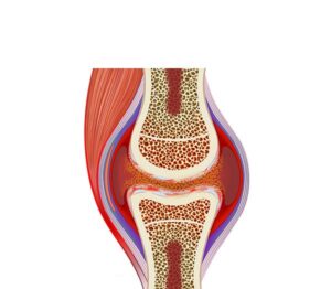
Synovial inflammation arthritis
24,50 € Add to basketSynovial inflammation arthritis. This illustration shows the process
where the membrane lining the joints presents evident swelling.
The synovial tissue thickens due to rheumatoid arthritis or physical
wear, producing chronic pain symptoms and a significant loss of
joint mobility. -
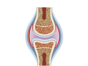
Synovial joint
24,50 € Add to basketSynovial joint. It is characterized as a type of movable connection
between bones. These joints are distinguished by the presence
of a synovial cavity containing synovial fluid, which facilitates
smooth movement between the articular surfaces. -
Template
24,50 € Add to basketLorem Ipsum es simplemente el texto de relleno de las imprentas
y archivos de texto. Lorem Ipsum ha sido el texto de relleno estándar
de las industrias desde el año 1500, cuando un impresor (N. del T.
persona que se dedica a la imprenta) desconocido usó