Skeleton
Showing 37–48 of 134 results
-
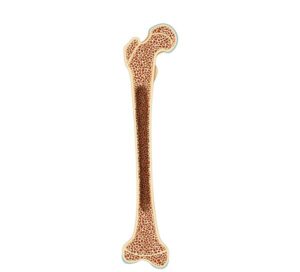
Internal section of the femur
24,50 € Add to basketThe cross-section of the femur provides a detailed illustration
of the internal anatomy of this thigh bone. Structures such as the
bone marrow, the femoral neck, the bone trabeculae, and the
distal and proximal epiphyses are highlighted.


-
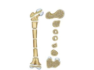
Internal structure of the femur
24,50 € Add to basketInternal structure of the femur, focusing on the walls that make up
its internal structure. Through cross-sectional views, it reveals the
different layers, thicknesses, cancellous bone, and the space occupied
by the bone marrow. The femoral neck and head are vital for the
hip joint. -
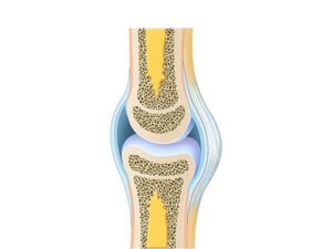
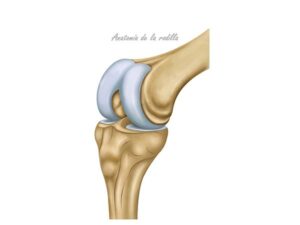
Knee and cartilage
24,50 € Add to basketThe knee and cartilage demonstrate the anatomical structure that
enables movement and shock absorption. The knee is composed
of the femur, tibia, and patella, along with the menisci and articular
cartilage, which protect the bones from wear and tear. -
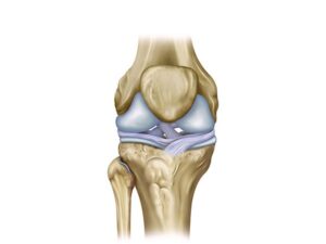
Knee cruciate ligament
24,50 € Add to basketKnee cruciate ligament. This anatomical structure joins the femur
with the tibia to provide internal stability. The anterior and posterior
cruciate ligaments are fundamental to preventing excessive bone
displacement, facilitating the diagnosis and treatment of common
joint injuries. -
Knee function
24,50 € Add to basketKnee function. This joint acts as a complex hinge that facilitates
the flexion and extension of the leg. Its structure, composed of the
femur and tibia, supports body weight and guarantees the stability
necessary to walk, run, and perform daily activities safely. -
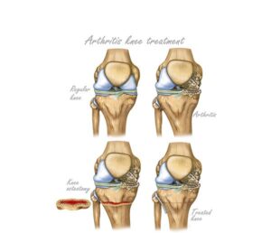
Knee osteotomy treatment
24,50 € Add to basketKnee osteotomy treatment is a surgical procedure that corrects
bone alignment to relieve pain and improve joint function. Moreover,
it redistributes load in early osteoarthritis and may delay the need for
knee replacement surgery. -
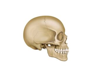
Lateral skull view
24,50 € Add to basketThe lateral skull shows frontal, parietal, and occipital bones,
protecting the brain and providing structural support and
facial shape. -
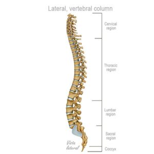
Lateral vertebral classification
24,50 € Add to basketLateral view of the vertebral column. This reveals a structure
composed of cervical (C1-C7), thoracic (T1-T12), lumbar (L1-L5),
and sacral (S1-S5) vertebrae, as well as the coccyx, which together
form the human spine. The natural curvature provides both support
and flexibility. -
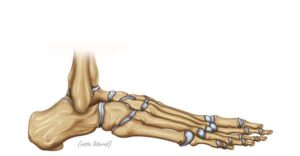
Lateral view of the foot
24,50 € Add to basketLateral view of the foot. Illustration of the bony anatomy showing
the connection of the tibia and fibula with the talus and calcaneus.
The image details the tarsal bones, such as the navicular and cuboid,
essential for maintaining balance, arch structure, and human mobility. -
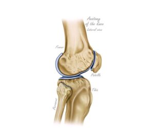
Lateral view of the knee
24,50 € Add to basketLateral view of the knee. This anatomical perspective details the
relationship between the femur, tibia, and patella. The illustration
highlights the articular surfaces and cartilages that allow for flexion
and extension of the leg, being fundamental for understanding
locomotion and skeletal stability. -
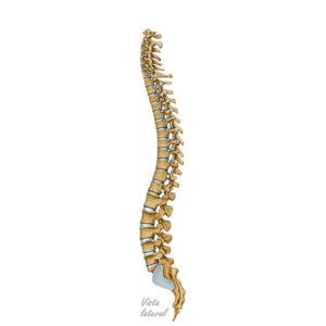
Lateral view of the spine
24,50 € Add to basketLateral view of the spine. It shows the segmented structure of the
vertebrae that make up the human back. It consists of cervical
vertebrae (C1–C7), thoracic vertebrae (T1–T12), lumbar vertebrae
(L1–L5), sacral vertebrae (S1–S5), and the coccyx. Its natural
curvature provides support and flexibility. -
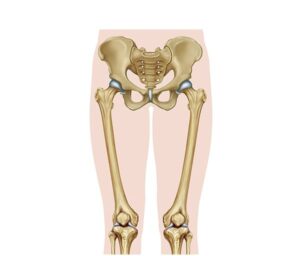
Lower limb anatomy
24,50 € Add to basketAnatomy of the lower limb. This is a complex biomechanical system
that influences the mobility and stability of the human body. The hip,
with the acetabulum and the femoral head, connects to the femur,
extending down to the knee.