Showing 37–48 of 1048 results
-
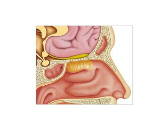
Anatomy and physiology of smell
24,50 € Add to basketAnatomy and physiology of smell: a system that detects odors
in the nasal mucosa, it transmits signals through the olfactory
nerve and processes them in the olfactory bulb in the brain.


-
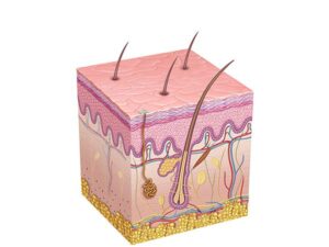
Anatomy and physiology of the skin
24,50 € Add to basketThe skin is the largest organ of the human body. It consists of
the epidermis, dermis, and hypodermis, which protect, regulate
temperature, and enable sensation through vessels, nerves, and
skin appendages. -
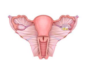
Anatomy and physiology of the uterus
24,50 € Add to basketAnatomy and physiology of the uterus: an organ composed of the
body, the isthmus, and the cervix that regulates the menstrual cycle
enables implantation, supports fetal development, and produces
contractions during childbirth. -
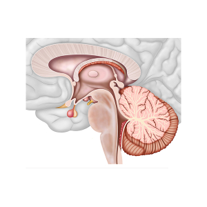
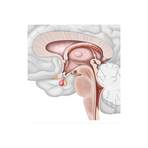
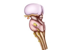
Anatomy Brainstem
24,50 € Add to basketThis illustration represents the anatomy of the
brainstem. First, it shows the inferior portion of
the brain, which connects the spinal cord with
higher structures. -
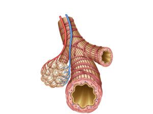
Anatomy of bronchioles and alveoli
24,50 € Add to basketBronchioles and alveoli are small respiratory structures
within the lungs. Bronchioles lack hyaline cartilage and
are surrounded by smooth muscle, while alveoli are tiny
air sacs where oxygen and carbon dioxide exchange occurs. -
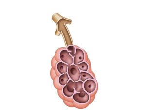
Anatomy of the alveolus
24,50 € Add to basketThe anatomy of the alveolus shows an air-filled cavity
with walls made of simple squamous epithelium. Elastic
and collagen fibers support pulmonary capillaries and
enable gas exchange. -
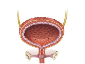
Anatomy of the Bladder
24,50 € Add to basketThe anatomy of the Bladder is a hollow muscular organ in the pelvis that stores urine. Supported by ligaments and connected to ureters and urethra, it allows controlled storage and elimination of urine.
-
Anatomy of the brain
24,50 € Add to basketThe illustration displays the brainstem. First,
this structure corresponds to the lower portion
of the brain. Additionally, it connects the spinal
cord with the upper structures of the brain. -
Anatomy of the brainstem
24,50 € Add to basketThe anatomy of the brainstem, also known as the brain
stem, is one of the most important structures of the central
nervous system. This region is located in the lower part of
the brain and establishes a direct connection between the
brain and the spinal cord..
-
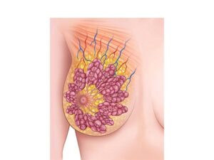
Anatomy of the breasts
24,50 € Add to basketThe anatomy of the breasts explains the internal organization
of breast tissue. It includes mammary lobes, lactiferous ducts,
adipose tissue, and a vascular network that supports the function
of the mammary glands. -
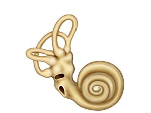
Anatomy of the cochlea
24,50 € Add to basketThe anatomy of the cochlea shows a spiral structure
in the inner ear that contains the organ of Corti. Hair
cells convert sound vibrations into electrical signals,
while fluid-filled chambers respond to different sound
frequencies. -
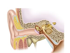
Anatomy of the ear
24,50 € Add to basketThe anatomy of the ear includes the outer, middle, and
inner ear. These structures work together to capture sound,
transmit vibrations, and maintain balance.