Marrow
Showing 109–120 of 183 results
-
Spinal cord anatomy
24,50 € Add to basketThe spinal cord is a crucial part of the central nervous
system. First, it extends from the brain to the interior
of the vertebral column. Additionally, it is composed
of nerves and specialized nervous tissue.


-
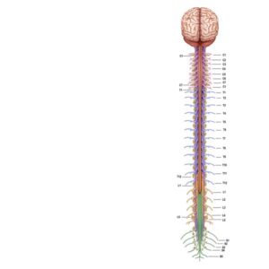
Spinal cord outline
24,50 € Add to basketA spinal nerve diagram illustrates the organization of these
important structures within the human nervous system.
Spinal nerves originate directly from the spinal cord and exit
through the intervertebral spaces of the vertebral column. -
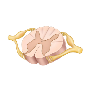
Spinal cord segment
24,50 € Add to basketThe spinal cord segment represents its
anatomical structure, including gray and
white matter, spinal nerves, and nerve roots.
This structure is fundamental for the
transmission of signals between the brain
and the body. -
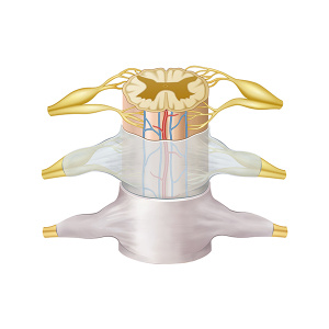
Spinal cord structure
24,50 € Add to basketThe structure of the spinal cord shows its segmented
organization and main components. First, it includes
the gray matter, which contains neuronal cell bodies.
-
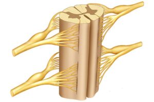
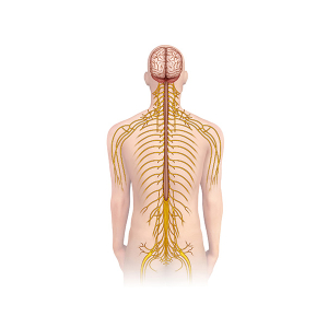
Spinal nerve branches
24,50 € Add to basketSpinal nerve branches play a fundamental role in the
organization of the human nervous system. These
nerves emerge directly from the spinal cord through
the intervertebral spaces of the vertebral column. -
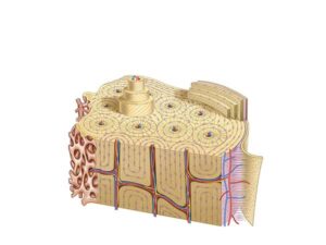
Spongy bone structure
24,50 € Add to basketStructure of cancellous bone. Also known as trabecular bone
tissue, it forms the inner part of bones and is characterized by a
porous, network-like structure. It consists of bone trabeculae
arranged irregularly, creating spaces filled with bone marrow. -
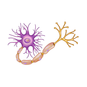
Structure of the Neuron
24,50 € Add to basketThe structure of the neuron is composed of three
main parts: the soma, the dendrites, and the axon.
The soma or cell body contains the nucleus and
regulates the essential functions of the cell. -
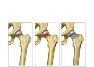
Subcapital femoral prosthesis
24,50 € Add to basketSubcapital femoral prosthesis is a surgical treatment for
severe fractures near the femoral head. Moreover, it replaces
damaged structures, relieving pain and restoring mobility.
Indication depends on patient age, health, and fracture severity. -
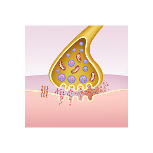
Synapse beta-amyloid protein
24,50 € Add to basketSynapse, beta-amyloid protein, and neurofibrillary tangles
in Alzheimer’s disease. The neuronal synapse is
the point of communication between neurons and plays
an essential role in the functioning of the nervous system..
-
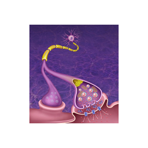
Synaptic Schema
24,50 € Add to basketThis synaptic schema visually represents the
functional connection between two neurons during
the process of neuronal communication. -
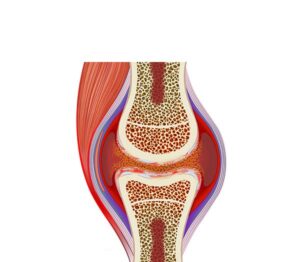
Synovial inflammation arthritis
24,50 € Add to basketSynovial inflammation arthritis. This illustration shows the process
where the membrane lining the joints presents evident swelling.
The synovial tissue thickens due to rheumatoid arthritis or physical
wear, producing chronic pain symptoms and a significant loss of
joint mobility. -
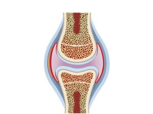
Synovial joint
24,50 € Add to basketSynovial joint. It is characterized as a type of movable connection
between bones. These joints are distinguished by the presence
of a synovial cavity containing synovial fluid, which facilitates
smooth movement between the articular surfaces.