Marrow
Showing 85–96 of 183 results
-
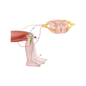
Myotatic reflex
24,50 € Add to basketThe myotatic reflex illustrates the neuromuscular
mechanism that enables involuntary muscle
contraction in response to stretching. This reflex
is essential for maintaining muscle tone and posture.


-
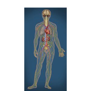
Nervous and circulatory systems
24,50 € Add to basketThe nervous system and the circulatory system are two
essential systems that support the proper functioning of
the human body. Both systems play important roles in
maintaining internal balance and ensuring that organs. -
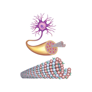
Neurofibrillary tangles
24,50 € Add to basketNeurofibrillary tangles are abnormal structures that
develop inside neurons. They are associated with
neurodegenerative diseases, such as Alzheimer’s disease.
-
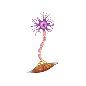
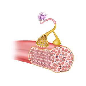
Neuromuscular junction
24,50 € Add to basketThe neuromuscular junction is the point where a
motor neuron and a muscle fiber interact. The
connection facilitates the transmission of signals
that initiate muscle contraction.
-
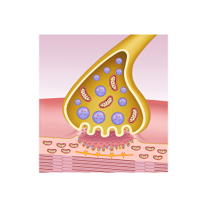
Neuromuscular synapse
24,50 € Add to basketThe neuromuscular synapse is the functional connection
between a motor neuron and a muscle fiber. It allows the
transmission of nerve signals that initiate muscle contraction. -
Neuromuscular synapse
24,50 € Add to basketThe neuromuscular synapse is the site of
communication between a motor neuron and
a muscle fiber. This junction plays a fundamental
role in muscle contraction. -
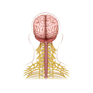
Organization of the cervical nerves
24,50 € Add to basketThe cervical nerve branch is the network of nerves
that originates in the spinal cord in the cervical
region of the spine. These nerves are distributed to
the neck, shoulders, and other nearby structures. -
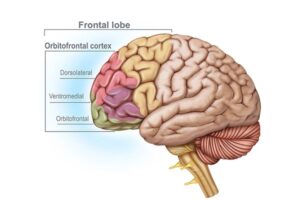
Physiology of the frontal lobe
24,50 € Add to basketIn frontal lobe physiology, we will see that it is one of
the most important regions of the human brain and
plays a fundamental role in numerous cognitive and
motor functions..
-
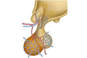
Pituitary gland
24,50 € Add to basketThe pituitary gland is a small endocrine gland
located at the base of the brain. Moreover, it plays
a central role in regulating the endocrine system. -
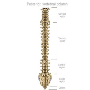
Posterior vertebral organization
24,50 € Add to basketPosterior view of the vertebral structure, highlighting its essential
function for mobility. Composed of cervical (C1-C7), thoracic (T1-T12),
lumbar (L1-L5), and sacral (S1-S5) vertebrae, and the coccyx, this
structure extends from the nape of the neck to the base of the back. -
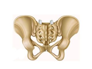
Posterior view of the hip
24,50 € Add to basketPosterior view of the hip. This vital joint connects the femur
to the sacrum, a triangular bone at the base of the spine. This
complex connection allows essential movements such as flexion
and rotation. -
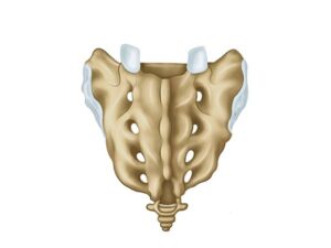
Posterior view of the sacrum
24,50 € Add to basketPosterior view of the sacrum: the triangular region at the bottom
of the spine. Numbered S1-S5, it features the median sacral crest,
intermediate and lateral crests, and four pairs of posterior sacral
foramina.