Marrow
Showing 49–60 of 183 results
-
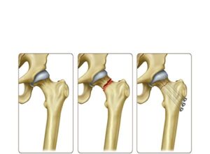
Femoral neck fracture treatment
24,50 € Add to basketFemoral neck fracture treatment involves internal fixation using
metal screws or nails to stabilize the bone and promote healing.
Moreover, it restores hip function. Treatment choice depends on
fracture severity and patient condition.


-
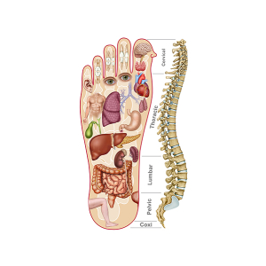
Foot reflexology
24,50 € Add to basketReflexology of the feet represents a map of the
soles where reflex zones connected to different
organs and body systems are indicated.
-
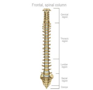
Frontal organization of the spine
24,50 € Add to basketThe frontal vertebral column highlights its crucial role as the
foundation for the mobility and stability of the human body.
It is composed of cervical (C1-C7), thoracic (T1-T12), and lumbar
(L1-L5), and sacral (S1-S5) vertebrae, as well as the coccyx. -
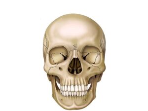
Frontal skull view
24,50 € Add to basketFrontal View of the Skull. In this view, the skull reveals the complexity
of its bony structure, which forms the facial anatomy and protects the
brain. Cranial bones such as the frontal and maxillary bones define
the facial appearance. -
Frontal view of the brain
24,50 € Add to basketFrontal view illustration of the brain. This shows
the right and left hemispheres, divided by the
interhemispheric fissure, a structure that separates
both halves, and allows their anatomical symmetry
to be appreciated. -
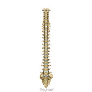
Frontal view of the spine
24,50 € Add to basketThe spinal column. It highlights its importance as a fundamental
pillar for the body’s mobility and stability. It consists of the cervical
vertebrae (C1–C7), thoracic vertebrae (T1–T12), lumbar vertebrae
(L1–L5), sacral vertebrae (S1–S5), and the coccyx. -
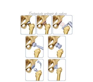
Hip osteoarthritis treatment
24,50 € Add to basketHip osteoarthritis and femoral neck treatment includes
physical therapy, medication, and prosthetic surgery. Moreover,
it aims to relieve pain and improve mobility. Treatment choice
depends on age, health status, and severity of joint damage. -
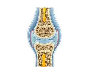
Human knee diagram
24,50 € Add to basketThe human knee. It displays its complex anatomical structure,
composed of bones, ligaments, cartilage, and muscles. The knee is
a vital joint that enables movements such as flexion and extension. -
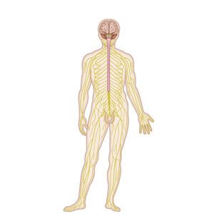
Human nervous system
24,50 € Add to basketThe human nervous system is a highly specialized network of
tissues that coordinates and regulates the functions of the body.
This system enables the organism to receive information from
the environment, process it, and generate appropriate responses. -
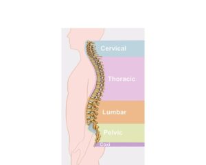
Human spinal classification
24,50 € Add to basketThe human vertebral classification consists of a
fundamental bone structure that provides support,
stability, and mobility to the body. They are organized
into: cervical, thoracic, lumbar, sacral, and coccygeal. -
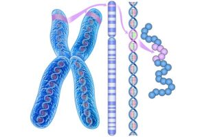
Huntington’s disease
24,50 € Add to basketHuntington’s disease (HD) is a hereditary neurodegenerative
disorder that progressively affects the nervous system. This
disease is caused by a mutation in the HTT gene, located on
chromosome 4..
-
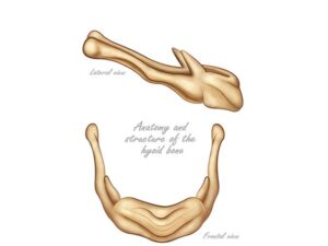
Hyoid bone
24,50 € Add to basketThe hyoid bone. It is a U-shaped structure located at the base
of the neck that does not articulate with other bones. It is situated
between the jaw and the larynx and is essential for functions such
as swallowing and speech.