Marrow
Showing 73–84 of 183 results
-
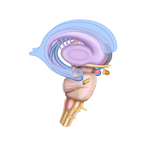
Limbic system anatomy
24,50 € Add to basketLimbic system anatomy comprises a set of brain
structures involved in regulating emotions, memory,
and motivation. First, this system includes the
hippocampus, the amygdala, and the hypothalamus.



-
Limbic system anatomy
24,50 € Add to basketLimbic system anatomy comprises a set of brain
structures involved in regulating emotions, memory,
and motivation. First, this system includes the
hippocampus, the amygdala, and the hypothalamus. -
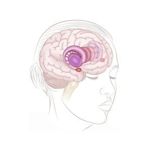
Limbic system and brain
24,50 € Add to basketRepresentation of the human head and the limbic
system shows the location of these structures
within the brain and key components such as the
hippocampus, amygdala, and hypothalamus. -
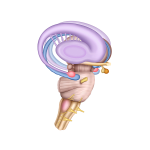
Limbic system structure
24,50 € Add to basketThe structure of the limbic system comprises
a set of interconnected brain regions. These
structures play a key role in emotions, memory,
and behavior. -
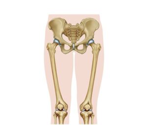
Lower limb anatomy
24,50 € Add to basketAnatomy of the lower limb. This is a complex biomechanical system
that influences the mobility and stability of the human body. The hip,
with the acetabulum and the femoral head, connects to the femur,
extending down to the knee. -
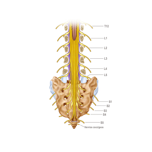
Lumbar and sacral nerves
24,50 € Add to basketThe lumbar and sacral nerves originate in the
spinal cord of the lumbar region of the spine and
are part of the peripheral nervous system. They play
a crucial role in transmitting signals between the
central nervous system -
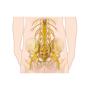
Lumbar spinal nerves
24,50 € Add to basketThe lumbar spinal nerves originate in the spinal cord
in the lumbar region of the spine. They are part of the
peripheral nervous system plays a crucial role in
transmitting signals between the central nervous system
and the lower extremities. -
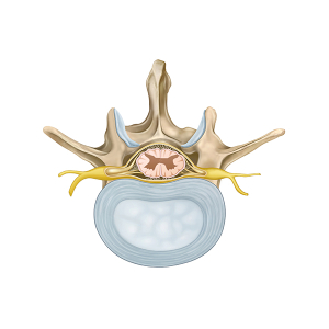
Lumbar vertebra anatomy
24,50 € Add to basketThe anatomy of the lumbar vertebra shows its robust
structure, designed to support the weight of the body
and allow flexion, extension, and rotation movements. -
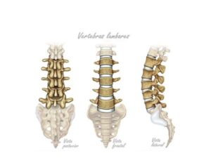
Lumbar vertebrae
24,50 € Add to basketLumbar vertebrae. The bony structures in the lower region of
the spine are shown here. These vertebrae, numbered from L1
to L5, support the weight of the torso and allow for movements
such as flexion and extension. -
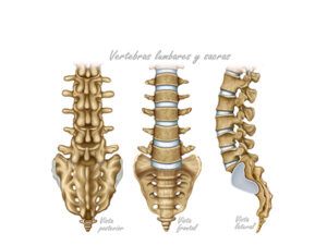
Lumbar vertebrae and sacrum
24,50 € Add to basketLumbar and sacral vertebrae. These show the bony structures
in the lower part of the spine. Numbered L1 through L5, the
lumbar vertebrae support the weight of the torso and allow for movements
such as flexion and extension. The sacrum, formed by fused vertebrae,
connects the lumbar spine to the pelvis.</p> -
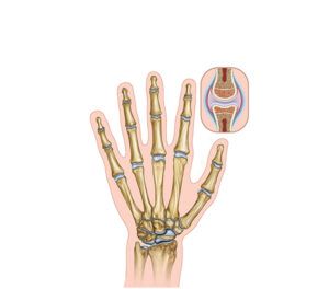
Morphology of the hand
24,50 € Add to basketAnatomy of the hand. This is a complex bony structure
that includes metacarpals, phalanges, and carpal bones.
The metacarpals form the base of the fingers, while the
phalanges make up the fingers themselves. -
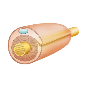
Myelin Sheath Structure
24,50 € Add to basketThe structure of the myelin sheath is a protective layer
composed mainly of lipids. It surrounds the axons of
nerve cells. In addition, it facilitates the transmission
of electrical signals.