Nerves
Showing 121–132 of 251 results
-
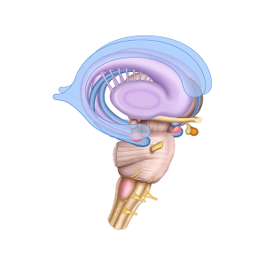
Limbic system anatomy
24,50 € Add to basketLimbic system anatomy comprises a set of brain
structures involved in regulating emotions, memory,
and motivation. First, this system includes the
hippocampus, the amygdala, and the hypothalamus.


-
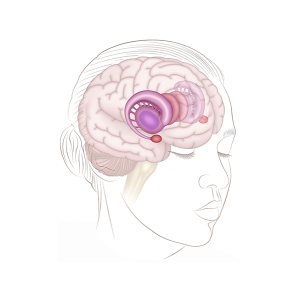
Limbic system and brain
24,50 € Add to basketRepresentation of the human head and the limbic
system shows the location of these structures
within the brain and key components such as the
hippocampus, amygdala, and hypothalamus. -
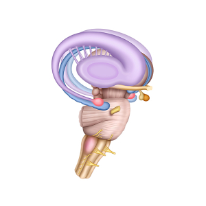
Limbic system structure
24,50 € Add to basketThe structure of the limbic system comprises
a set of interconnected brain regions. These
structures play a key role in emotions, memory,
and behavior. -
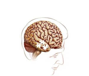
Lost memories in Alzheimer’s disease
24,50 € Add to basketThe lost memories in Alzheimer’s disease have an altered function in
cognitive processes. Moreover, under normal conditions, neurons
communicate efficiently. Additionally, they store and retrieve information. -
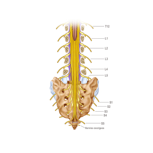
Lumbar and sacral nerves
24,50 € Add to basketThe lumbar and sacral nerves originate in the
spinal cord of the lumbar region of the spine and
are part of the peripheral nervous system. They play
a crucial role in transmitting signals between the
central nervous system -
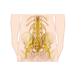
Lumbar spinal nerves
24,50 € Add to basketThe lumbar spinal nerves originate in the spinal cord
in the lumbar region of the spine. They are part of the
peripheral nervous system plays a crucial role in
transmitting signals between the central nervous system
and the lower extremities. -
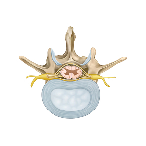
Lumbar vertebra anatomy
24,50 € Add to basketThe anatomy of the lumbar vertebra shows its robust
structure, designed to support the weight of the body
and allow flexion, extension, and rotation movements. -
Macula and fovea
24,50 € Add to basketThe macula and fovea are central retinal regions responsible
for detailed vision. The macula supports high visual acuity,
while the fovea contains the highest density of cone cells,
allowing precise color perception and sharp central vision. -
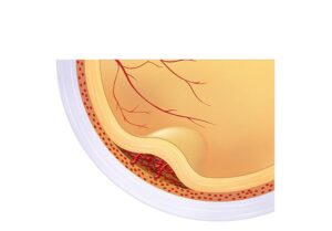
Macular Degeneration
24,50 € Add to basketMacular Degeneration (AMD) illustrates how the condition affects
the macula, the central part of the retina responsible for detailed
vision. Aging can damage the cells of the macula, leading to blurred
vision or loss of central vision. -
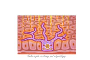
Melanocyte physiology
24,50 € Add to basketEs un hecho establecido hace demasiado tiempo que
un lector se distraerá con el contenido del texto de un
sitio mientras que mira su diseño. El punto de usar
Lorem Ipsum -
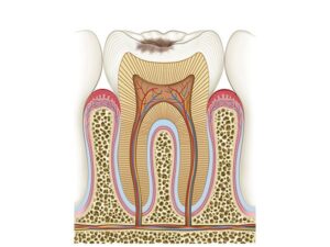
Molar caries stage II
24,50 € Add to basketThe molar caries stage II has an altered function in tooth integrity.
Moreover, under normal conditions, enamel protects the dentin.
Additionally, dentin transmits stimuli to the pulp. Furthermore,
these structures maintain dental function. -
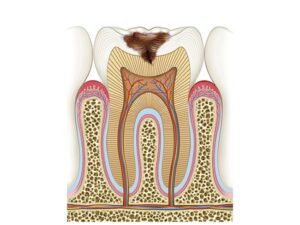
Molar caries stage III
24,50 € Add to basketThe molar caries stage III has an altered function in tooth integrity.
Moreover, under normal conditions, enamel protects the inner layers.
Additionally, dentin provides structural support. Furthermore, they
ensure proper dental function.