Nerves
Showing 217–228 of 251 results
-
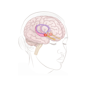
The brain tonsils
24,50 € Add to basketThe brain tonsils. This representation of the human
head shows the location and function of these key
structures in the brain. First, the amygdalae are
located in the temporal lobe.


-
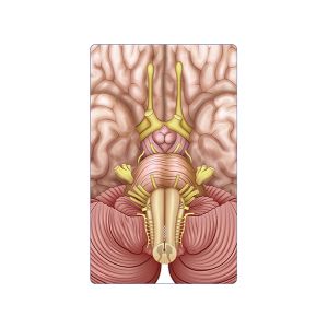
The brainstem
24,50 € Add to basketThe brainstem provides a detailed view of the
structures in the lower part of the encephalon.
First, it regulates vital functions such as breathing
and heart rate.
-
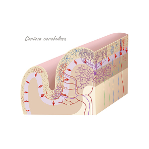
The cerebellar cortex
24,50 € Add to basketThis illustration shows the cerebellar cortex, a
structure essential for motor coordination. First,
it is important to note that it is organized into
three well-defined layers. -
The cerebral amygdala
24,50 € Add to basketThe amygdala is a brain structure located in the
temporal lobe. First, it is essential for processing
emotions, especially fear and aggression. -
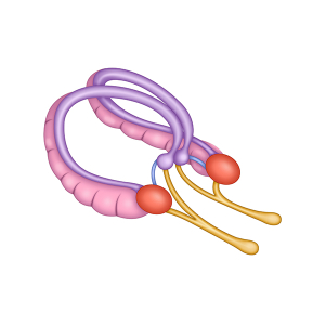
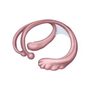
The cerebral fornix
24,50 € Add to basketThe structure of the cerebral fornix constitutes
a pathway of nerve fibers that connects various
regions of the brain. Involved in areas of
memory, emotions, and communication. -
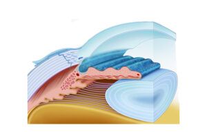
The cornea and iris
24,50 € Add to basketThe cornea and iris are anterior eye structures that control
light entry and help focus images on the retina. The cornea
refracts light while the iris regulates pupil size to adapt vision
to different lighting conditions. -
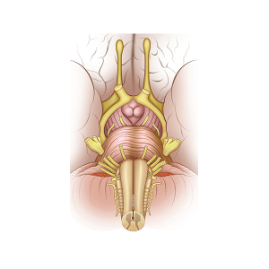
The cranial nerves
24,50 € Add to basketThe cranial nerves are a key part of the nervous system.
First, they connect the brain to the spinal cord and
control vital functions. Additionally, they contain
nuclei that regulate sensory and motor functions
of the head and neck. -
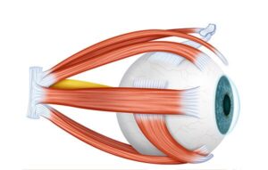
The eye muscles
24,50 € Add to basketOcular muscles control eye position and movement. Six
muscles work together to produce vertical, horizontal, and
rotational movements, allowing coordinated gaze and stable
visual tracking. -
The facial nerves
24,50 € Add to basketThe facial nerves are shown on a female face, where
their distribution and function are clearly highlighted.
First, they correspond to cranial nerve VII. -
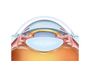
The glaucoma
24,50 € Add to basketOcular glaucoma represents a condition characterized by an increase
in intraocular pressure, which can damage the optic nerve and cause
vision loss. The accumulation of aqueous humor due to inefficient
drainage is shown. -
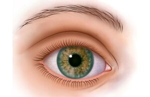
The human eye
24,50 € Add to basketThe external anatomy of the eye includes the cornea,
sclera, iris, pupil, and eyelids that protect the eye and
regulate the entry of light. -
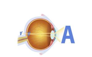
The hyperopia vision
24,50 € Add to basketFarsighted vision, or distance vision, is an eye condition where
nearby objects appear blurry because the image forms behind
the retina. This usually happens due to a shorter eye or a flatter
cornea.