Nerves
Showing 61–72 of 251 results
-
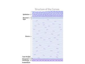
Cornea and ocular surface
24,50 € Add to basketThe cornea and ocular surface allow light to enter the eye
and contribute to focusing images on the retina. In addition,
the cornea protects the eye and maintains visual clarity
through its transparency and curved structure.


-
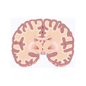
Coronal brain section
24,50 € Add to basketSchematic illustration of the coronal section
of the brain. In this view, several important
internal structures can be clearly observed. -
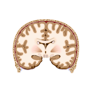
Coronal section of the brain
24,50 € Add to basketThis anatomical illustration of a coronal section
of the brain shows the brain structure in detail.
First, it highlights key elements such as the
cortex, hypothalamus, and ventricles. -
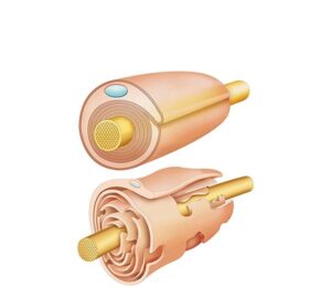
Demyelinating disease
24,50 € Add to basketThe demyelinating disease has an altered function in nervous system
transmission. Moreover, under normal conditions, myelin acts as an
insulator. Additionally, it allows rapid conduction of nerve impulses.
Furthermore, it ensures efficient neuronal communication. -
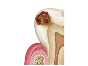
Dental caries
24,50 € Add to basketThe dental caries have an altered function in tooth integrity.
Moreover, under normal conditions, enamel protects the inner
layers. Additionally, dentin provides structural support.
Furthermore, they maintain dental health. -
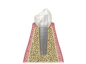
Dental implant diagram
24,50 € Add to basketThe dental implant diagram has an essential function in oral
rehabilitation. Moreover, under normal conditions, tooth roots
anchor teeth to the bone. Additionally, they support chewing
forces. Furthermore, they ensure dental stability and function. -
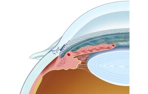
Detail of Schlemm’s canal
24,50 € Add to basketSchlemm’s canal collects aqueous humor filtered by the
trabecular meshwork and directs it to the venous system,
helping regulate eye pressure. -
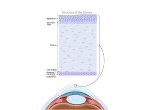
Diagram of the cornea
24,50 € Add to basketThe corneal layer is the transparent front structure of
the eye. It allows light to enter the eye, contributes to
light refraction, and protects internal eye structures,
helping form clear images on the retina. -
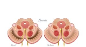
Dopamine and substantia nigra
24,50 € Add to basketThe dopamine and the substantia nigra have an essential function
in the nervous system. Moreover, dopamine regulates voluntary
movements. Additionally, it plays a role in mood and motivation. -
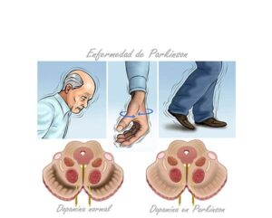
Dopamine in Parkinson’s disease
24,50 € Add to basketDopamine is a neurotransmitter and plays a crucial role in the
regulation of movement and mood. This deficiency of dopamine
leads to motor and non-motor symptoms, such as problems with
balance and cognitive alterations. -
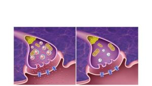
Dopaminergic synapse
24,50 € Add to basketThe dopaminergic synapse has an essential function in neuronal
transmission. Moreover, dopamine acts as a key neurotransmitter.
Additionally, it facilitates communication between neurons. -
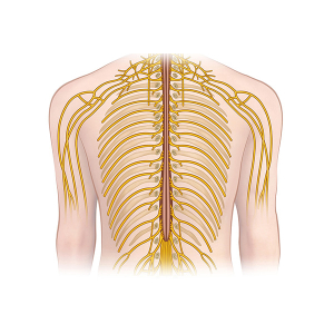
Dorsal nerve branching
24,50 € Add to basketThe dorsal nerve branching refers to the extension of
nerves from the posterior part of the spinal cord. This
structure enables the transmission of sensory information
to the central nervous system.