Showing 193–204 of 1048 results
-
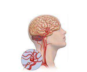
Cerebral arterial aneurysm
24,50 € Add to basketCerebral arterial aneurysm. Detailed medical illustration of a
human head, where weakened bulges in the artery are visualized.
This condition shows how vessels fill with blood, pressing on
surrounding tissue and creating a high risk of severe hemorrhage
in the patient.


-
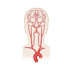
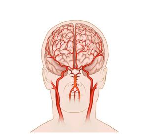
Cerebral arterial irrigation
24,50 € Add to basketCerebral arterial irrigation. This illustration describes the supply
of oxygen-rich blood through the main arteries to the brain. The
diagram shows how the internal carotids and vertebrals nourish the
tissue, being a vital process for maintaining healthy neurological function. -
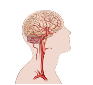
Cerebral blood circulation
24,50 € Add to basketCerebral blood circulation. This illustration represents the flow
of oxygenated blood from the arteries to the capillaries for nutrient
exchange. The graphic shows how veins return deoxygenated blood
to the heart, facilitating the understanding of cellular perfusion. -
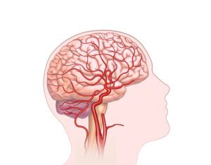
Cerebral blood flow
24,50 € Add to basketLorem Ipsum es simplemente el texto de relleno de las imprentas
y archivos de texto. Lorem Ipsum ha sido el texto de relleno estándar
de las industrias desde el año 1500, cuando un impresor (N. del T.
persona que se dedica a la imprenta) desconocido usó -
Cerebral carotid irrigation
24,50 € Add to basketCerebral carotid irrigation. This illustration shows the blood
supply through the internal carotids and vertebral arteries connected
by the Circle of Willis. The diagram details how the internal carotid
nourishes the forebrain and the upper lobes for cognitive function. -
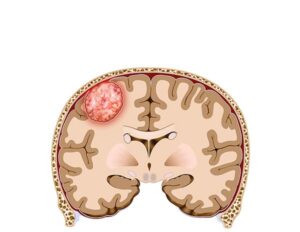
Cerebral meningioma tumor
24,50 € Add to basketCerebral meningioma tumor. This realistic illustration details
a tumor originating in the meningeal membranes and its impact on
brain structure. The content highlights the importance of this visual
resource for clinical diagnosis, surgical planning, and education. -
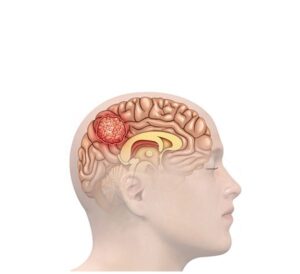
Cerebral meningioma tumor
24,50 € Add to basketCerebral meningioma tumor. This realistic illustration details
a tumor originating in the meningeal membranes and its impact on
brain structure. The content highlights the importance of this visual
resource for clinical diagnosis, surgical planning, and education. -
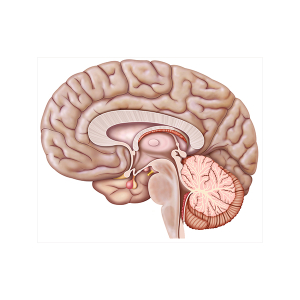
Cerebral sagittal section
24,50 € Add to basketIllustration of the sagittal section of the encephalon.
This view provides a detailed anatomical perspective
of the brain’s structure in profile, allowing observation
of the internal arrangement of its main components. -
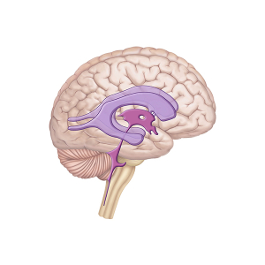
Cerebrospinal fluid
24,50 € Add to basketThis representation schematically illustrates cerebrospinal
fluid (CSF). First, it highlights the fundamental role of CSF
in protecting and nourishing the central nervous system. -
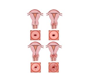
Cervical cancer stages
24,50 € Add to basketCervical cancer stages describe tumor progression from localized
abnormal cells to spread into nearby and distant organs. This
classification is essential for guiding treatment and determining
prognosis. -
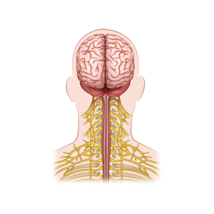
Cervical nerve branch
24,50 € Add to basketThe cervical nerve branch is the network of nerves
that originates in the spinal cord in the cervical
region of the spine. These nerves are distributed to
the neck, shoulders, and other nearby structures. -
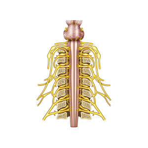
Cervical nerve structure
24,50 € Add to basketThe classification of the cervical nerves describes the
organization of the nerves that originate in the cervical
region of the spinal cord. These nerves are distributed to
the neck, shoulders, and other nearby structures.