Showing 529–540 of 1048 results
-
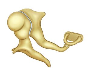
Middle ear bones
24,50 € Add to basketMiddle ear ossicles, the malleus, incus, and stapes,
transmit and amplify sound vibrations from the eardrum
to the inner ear. This process allows sound waves to be
converted into electrical signals that the brain interprets
as sound.


-
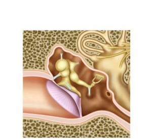
Middle ear structure
24,50 € Add to basketThe middle ear is located in the tympanic cavity and
contains the eardrum and auditory ossicles. These
structures transmit and amplify sound vibrations toward
the inner ear, allowing efficient hearing and proper
sound processing. -
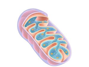
Mitochondria anatomy and physiology
24,50 € Add to basketLa mitocondria es un orgánulo con doble membrana que
produce ATP mediante la cadena respiratoria, siendo clave
en el metabolismo celular y la homeostasis energética. -
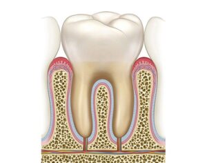
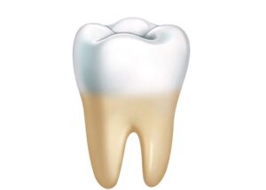
Molar and gingival structure
24,50 € Add to basketMolars are posterior teeth specialized for grinding food.
Their structure includes enamel, dentin, pulp, and roots
anchored to the jaw by the periodontal ligament. -
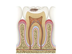
Molar caries stage II
24,50 € Add to basketThe molar caries stage II has an altered function in tooth integrity.
Moreover, under normal conditions, enamel protects the dentin.
Additionally, dentin transmits stimuli to the pulp. Furthermore,
these structures maintain dental function. -
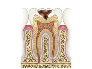
Molar caries stage III
24,50 € Add to basketThe molar caries stage III has an altered function in tooth integrity.
Moreover, under normal conditions, enamel protects the inner layers.
Additionally, dentin provides structural support. Furthermore, they
ensure proper dental function. -
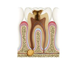
Molar caries stage IV
24,50 € Add to basketThe molar caries stage IV has a severely altered function in tooth
integrity. Moreover, under normal conditions, enamel and dentin
protect the pulp. Additionally, the pulp contains nerves and blood
vessels. -
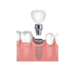
Molar implant
24,50 € Add to basketThe molar implant has an essential function in dental rehabilitation.
Moreover, under normal conditions, molars allow food grinding.
Additionally, they contribute to dental arch stability. Furthermore,
they ensure efficient chewing. -
Molar tooth anatomy
24,50 € Add to basketMolar tooth anatomy is a posterior tooth specialized in
grinding food. Its structure includes enamel, dentin,
and pulp, as well as roots that attach it to the bone
via the periodontal ligament. -
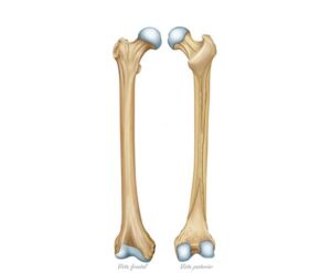
Morphology of the femur bone
24,50 € Add to basketMorphology of the femur bone. The longest bone in the thigh
presents a robust structure with a well-defined head, neck, and body.
Its frontal and posterior views reveal details such as the condyles and
the intertrochanteric crest, crucial elements for body support and
joint connection. -
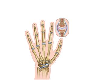
Morphology of the hand
24,50 € Add to basketAnatomy of the hand. This is a complex bony structure
that includes metacarpals, phalanges, and carpal bones.
The metacarpals form the base of the fingers, while the
phalanges make up the fingers themselves. -
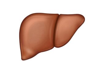
Morphology of the liver
24,50 € Add to basketThe morphology of the liver shows an organ located
in the upper right abdomen, composed of hepatic
lobes and hepatocytes that perform metabolic, digestive,
and detoxification functions.