Showing 469–480 of 1048 results
-
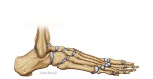
Lateral view of the foot
24,50 € Add to basketLateral view of the foot. Illustration of the bony anatomy showing
the connection of the tibia and fibula with the talus and calcaneus.
The image details the tarsal bones, such as the navicular and cuboid,
essential for maintaining balance, arch structure, and human mobility.


-
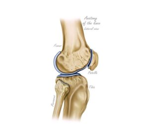
Lateral view of the knee
24,50 € Add to basketLateral view of the knee. This anatomical perspective details the
relationship between the femur, tibia, and patella. The illustration
highlights the articular surfaces and cartilages that allow for flexion
and extension of the leg, being fundamental for understanding
locomotion and skeletal stability. -
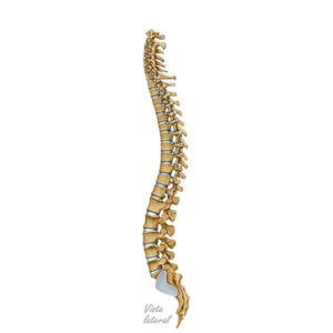
Lateral view of the spine
24,50 € Add to basketLateral view of the spine. It shows the segmented structure of the
vertebrae that make up the human back. It consists of cervical
vertebrae (C1–C7), thoracic vertebrae (T1–T12), lumbar vertebrae
(L1–L5), sacral vertebrae (S1–S5), and the coccyx. Its natural
curvature provides support and flexibility. -
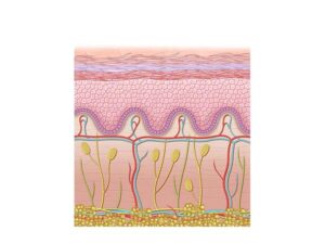
Layers of human skin
24,50 € Add to basketHuman skin is composed of epidermis, dermis, and hypodermis.
These layers protect against pathogens, regulate temperature,
and enable sensory perception through melanin, vessels, and skin
appendages. -
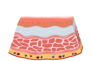
Layers of the bladder wall
24,50 € Add to basketThe bladder wall consists of mucosa, submucosa, detrusor
muscle, and serosa. These layers allow expansion, storage,
and contraction of the bladder during urination. -
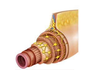
Layers of the digestive tract
24,50 € Add to basketThe layers of the digestive tract include mucosa, submucosa,
muscular layer, and serosa. These structures support digestion,
enzyme secretion, nutrient absorption, and movement of food
through the system. -
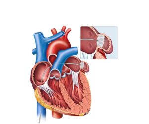
Left atrial appendage
24,50 € Add to basketThe left atrial appendage has a general function related to atrial
dynamics. Moreover, under normal conditions, it contributes to
left atrial contraction. Additionally, it may act as a blood reservoir. -
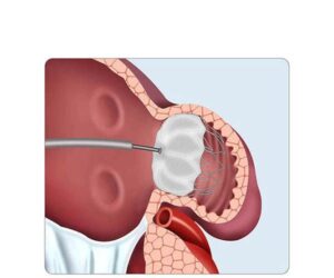
Left atrial appendage closure
24,50 € Add to basketThe left atrial appendage closure has a general preventive
function in the cardiovascular system. Moreover, under normal
conditions, the appendage contributes to atrial dynamics.
Additionally, it may act as a blood reservoir. -
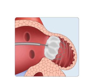
Left atrial appendage closure
24,50 € Add to basketThe left atrial appendage closure has a general preventive function
in the cardiovascular system. Moreover, under normal conditions,
the appendage contributes to atrial dynamics. Additionally, it may
act as a blood reservoir. -
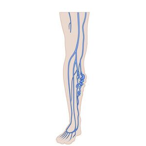
Leg with varicose veins
24,50 € Add to basketLeg with varicose veins shows dilated and twisted veins caused
by venous valve failure, leading to blood pooling, pain, swelling,
and visible skin changes. -
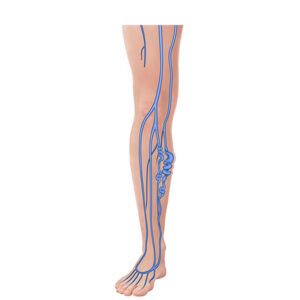
Legs affected by varicose veins
24,50 € Add to basketLegs affected by varicose veins show dilated, twisted veins caused
by venous insufficiency, leading to pain, swelling, and heaviness,
with potential complications if untreated. -
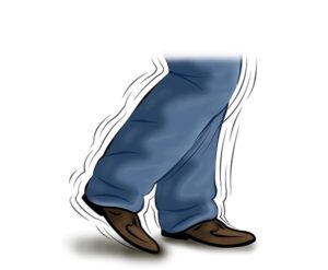
Legs in Parkinson’s disease
24,50 € Add to basketThe legs in Parkinson’s disease have an altered function in mobility.
Moreover, under normal conditions, dopamine regulates voluntary
movements. Additionally, it allows smooth and coordinated execution.